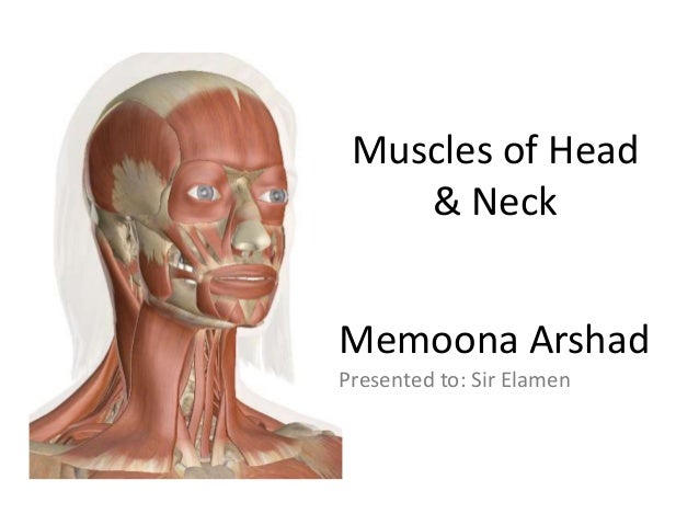
38 head and neck muscle diagram
Muscles of Head and Neck. Anterior and Lateral Neck Muscles Blank Diagram Complete Diagram Supra/Infrahyoid Muscles Blank Diagram Complete Diagram Pharyngeal Constrictor Muscles Blank Diagram Complete Diagram Muscles of Facial Expression Blank Diagram Complete Diagram Muscles of Mastication Lateral Skull (Blank Diagram) Mastication 1(Complete ...
Cancers that are known collectively as head and neck cancers usually begin in the squamous cells that line the mucosal surfaces of the head and neck (for example, those inside the mouth, throat, and voice box). These cancers are referred to as squamous cell carcinomas of the head and neck. Head and neck cancers can also begin in the salivary glands, sinuses, or muscles or nerves in the head ...
The anatomy of the head and neck of the human body, including the bones, muscles, blood vessels, nerves, glands, nose, mouth, and throat. - Head & Neck Anatomy 2019 - 3D model by INTERVOKE (@intervoke) [2500546]

Head and neck muscle diagram
Educational video describing the muscle anatomy of the neck. Become a friend on facebook: http://www.facebook.com/drebraheim Follow me on twitter: https://tw
April 25, 2013 - The origins of the muscles of facial expression are on the surface of the skull (remember, the origin of a muscle does not move). The insertions of thes...
Muscles of the Head and Neck. Humans have well-developed muscles in the face that permit a large variety of facial expressions. Because the muscles are used to show surprise, disgust, anger, fear, and other emotions, they are an important means of nonverbal communication. Muscles of facial expression include frontalis, orbicularis oris, laris ...
Head and neck muscle diagram.
28.10.2021 · Head and neck Thorax Abdomen and pelvis Upper limb Lower limb. Histology. General . Introduction to cells and tissues Epithelial tissue Connective tissue Nervous tissue Muscle tissue Cartilage and bone. Systems. Cardiovascular system Nervous system Integumentary system Musculoskeletal system Respiratory system Urinary system Endocrine …
The neck muscles, including the sternocleidomastoid and the trapezius, are responsible for the gross motor movement in the muscular system of the head and neck. They move the head in every direction, pulling the skull and jaw towards the shoulders, spine, and scapula. Working in pairs on the left and right sides of the body, these muscles ...
21.01.2018 · The neck is the start of the spinal column and spinal cord. The spinal column contains about two dozen inter-connected, oddly shaped, bony segments, called vertebrae. The neck contains seven of ...
The humerus (/ ˈ h j uː m ər ə s /, plural: humeri) is a long bone in the arm that runs from the shoulder to the elbow.It connects the scapula and the two bones of the lower arm, the radius and ulna, and consists of three sections.The humeral upper extremity consists of a rounded head, a narrow neck, and two short processes (tubercles, sometimes called tuberosities).
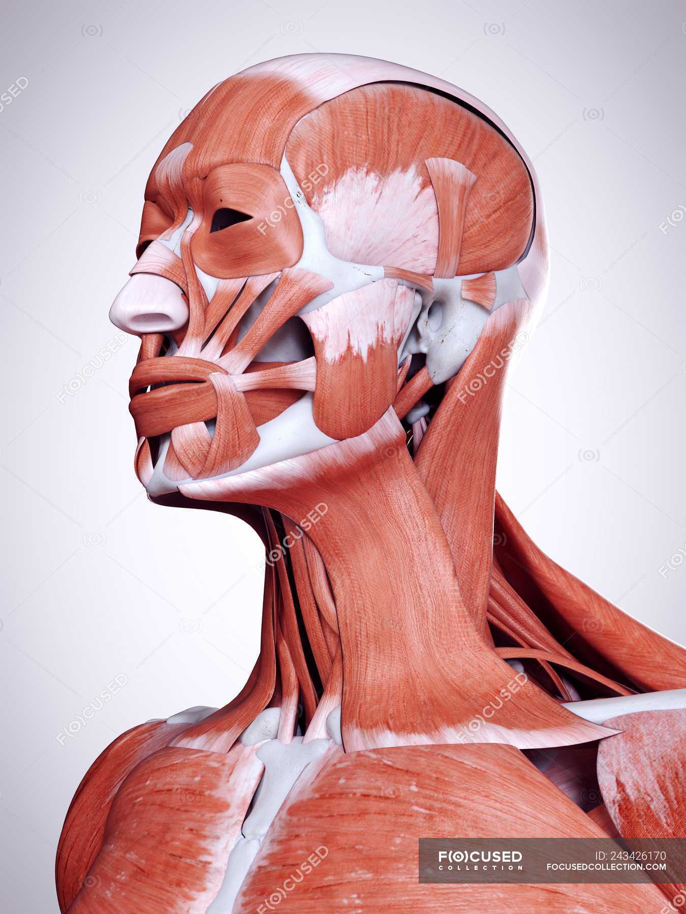
The muscle anatomy of the head and neck is a fascinating area, with the the neck also containing the 7 vertebrae of the part of the spine called the cervical curve. Superficial dissections of the head and neck as seen in the gallery, show the many different muscles that are required for movement plus those that control facial expression.
The primary muscles of mastication (chewing food) are the temporalis, medial pterygoid, lateral pterygoid, and masseter muscles. The four main muscles of mastication attach to the rami of the mandible and function to move the jaw (mandible). The cardinal mandibular movements of mastication are elevation, depression, protrusion, retraction, and side to side movement. To augment the process of ...
May 31, 2019 - The neck muscles and other soft tissues—such as ligaments and blood vessels—play important roles in the cervical spine’s movements, stability, and function.
April 24, 2020 - Superficial muscles are the muscles closest to the skin surface and can usually be seen while a body is performing actions. Many in the neck help to stabilize or move the head. Some also create facial expressions.
Lateral View - Head, Neck, and Shoulder Muscles . Lateral View - Pectoral Girdle The cleidocervicalis is labeled clavotrapezius in your book. This figure illustrates the position of the transversus abdominus in relation to the internal and external oblique muscles.
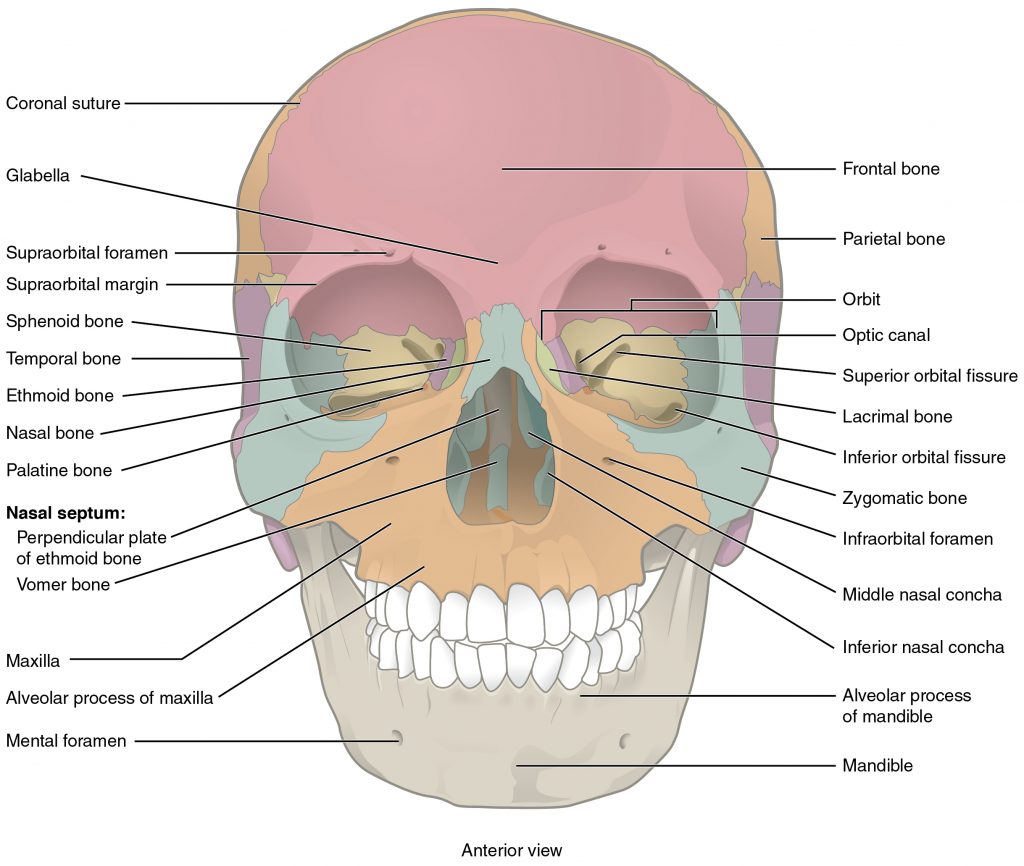
Skin. The head and neck is covered in skin and its appendages, termed the integumentary system.These include hair, sweat glands, sebaceous glands, and sensory nerves.The skin is made up of three microscopic layers: epidermis, dermis, and hypodermis.The epidermis is composed of stratified squamous epithelium and is divided into the following five sublayers or strata, listed in order from outer ...
December 4, 2018 - Anatomy of The Neck: Causes of Neck Pain and How to Manage the Pain In basic terms, the neck (cervical spine) joins the shoulders and chest to the head. The neck is one of the most complex and intricate structures in our body and includes the spinal cord, which sends messages from the brain ...
30.04.2021 · Examples of where lymph glands group together are the sides of the neck, the armpits and the groins. The diagram shows the main groups of lymph glands in the head and neck. However, lymph glands occur in many other places in the body. Lymph glands are joined together by a network of lymph channels. Lymph is a fluid that forms between the cells of the …
Muscles of the head and neck diagram | quizlet
DENT-1431: Head and Neck Anatomy 3 5. Online Continuing Education Assignment 6. Client Education Brochures Course Content Outline: 1. Skull a. Bones b. Sutures c. Foramens d. Fossae 2. Muscles of the head and neck a. Muscles of facial expression b. Muscles of mastication c. Hyoid muscles d. Muscles of the tongue and pharynx 3. Temporomandibular ...
Muscles of head and neck
13,402 anatomy of the head and neck stock photos, vectors, and illustrations are available royalty-free. See anatomy of the head and neck stock video clips. of 135. muscle head anatomy vocal organ diagram female neck anatomy neck wireframe head neck human anatomy head artery anatomy face pharynx vector neck degree head anatomy 3d.
7.2 head and neck basic concepts – nursing skills
The suboccipital muscles act to rotate the head and extend the neck. Rectus capitis posterior major and Rectus capitis posterior minor attach the inferior nuchal line of the occiput to the C2 and C1 vertebrae respectively. Obliquus capitis superior also extends from the occiput to C1 while ...
3d rendered illustration of head and neck muscles in human ...
The sternocleidomastoid is a large, two-headed muscle of the neck. Its clavicular head originates from the medial third of the clavicle, while its sternal head arises from the manubrium of sternum . The heads come together and ascend diagonally to insert onto the mastoid process of the temporal bone .

The muscles of the head and neck - stock image - f001/7815 ...
Neck muscles are bodies of tissue that produce motion in the neck when stimulated. The muscles of the neck run from the base of the skull to the upper back and work together to bend the head and ...
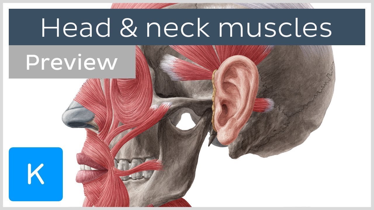
Main muscles of the head and neck (preview) - human anatomy | kenhub
Human muscle system, the muscles of the human body that work the skeletal system, that are under voluntary control, and that are concerned with movement, posture, and balance. Broadly considered, human muscle—like the muscles of all vertebrates—is often divided into striated muscle, smooth muscle, and cardiac muscle.

Anatomy of muscles on back of head stock illustration ...
Cross-sectional labeled anatomy of the head and neck of the domestic cat on CT imaging (bones of the skull, cervical spine, mandible, hyoid bone, muscles of the neck, nasal cavity and paranasal sinuses, oral cavity, larynx)
Muscles of the head, neck and back (illustrations ...
We are pleased to provide you with the picture named Back And Neck Muscles Diagram.We hope this picture Back And Neck Muscles Diagram can help you study and research. for more anatomy content please follow us and visit our website: www.anatomynote.com. Anatomynote.com found Back And Neck Muscles Diagram from plenty of anatomical pictures on the internet.

Muscular anatomy of the head and neck | neck muscle anatomy ...
16.09.2021 · The biceps muscle have “short head” and a “long head” that work as a single muscle. The origin at the scapula and the insertion into the radius of the biceps brachii mean it can act on both the shoulder joint and the elbow joint, which is why this muscle participates in a few movements of the arm. The main function of the biceps muscle is the pulls the forearm up …

Head and neck muscles - stock image - n150/0030 - science ...
The origin is the maxilla and mandible, the alveolar margins near the molars and the insertion is the orbicularis oris. ... This is a superficial muscle that tightens the neck, helps lower the jaw, helps draw down the lower lip and angle of the mouth during grimacing/melancholy expression as ...

Head and neck muscles diagram
5 Aug 2021 — Neck Muscle Anatomy ... The muscles of the neck have primary functions to stabilize and allow movement of the head, neck, and spine. They also ...What are the major muscles in the neck?How many muscles control the head and neck?

Muscles of the head & neck | anatomy model - youtube
Head and neck (anterior view) The head and neck are two examples of the perfect anatomical marriage between form and function, mixed with a dash of complexity. The neck is resilient enough to sustain a five kilogram weight 24/7, yet sufficiently mobile to move it in several directions.

Posterior triangle of the neck head and neck anatomy ...
Ninja nerds, In this video we discuss the muscles of the head & neck. Correction at 10:03 [#56]: It is the THYROHYOID, apologies for the mistake. ***PLEASE S...

Head and neck human anatomy (muscles)
Quizzes on the muscles of the head and neck. The quizzes below each include 15 multiple-choice identification questions related to the muscles of the head and neck. There are three sections for you to practice: muscle identification, muscle actions, and muscle origins and insertions.
Muscles of the face head and neck functions
CT scan of head and neck : Radiological anatomy of the head and neck on a CT in axial, coronal, and sagittal sections, and on a 3D images

Muscles of head and neck diagram diagram | quizlet
A single platysma muscle is only shown in the lateral view of the head muscles in Figure 8-4. There are two platysma muscles, one on each side of the neck. Each is a broad sheet of a muscle that covers most of the anterior neck on that side of the body. The other anterior neck muscles are below ...
Thoracic and head/neck muscles - ppt download
The neck is the bridge between the head and the rest of the body. It is located in between the mandible and the clavicle, connecting the head directly to the torso, and contains numerous vital structures. It contains some of the most complex and intricate anatomy in the body and is comprised of numerous organs and tissues with essential structure and function for normal physiology.

Muscles - head | neck muscle anatomy, human body anatomy ...
See Otolaryngol Head Neck Surg 2004 Nov;131(5):596-600 and Surgery 2004 Dec;136(6):1310-22. It important to consider that routine use of the nerve monitoring system provides valuabe experience and confidence in troubleshooting the problems that inevitably arise. To this end, the below troubleshooting guide has been developed using instructions provided by the device …

Homo sapiens neck thorax muscle organ, others, hand, human ...
Feb 22, 2012 - This Pin was discovered by Morgan Raimondo. Discover (and save!) your own Pins on Pinterest
Neck muscles anatomy: list, origins, insertions, action | kenhub
The superior oblique muscle, or obliquus oculi superior, is a fusiform muscle originating in the upper, medial side of the orbit (i.e. from beside the nose) which abducts, depresses and internally rotates the eye. It is the only extraocular muscle innervated by the trochlear nerve (the fourth cranial nerve). Structure. The superior oblique muscle loops through a pulley-like structure …
Head and neck laminated anatomical chart
July 6, 2020 - Nasalis: This muscle helps you scrunch your nose by compressing the bridge of the nose and pulling the nostrils open. Mentalis: This muscle causes wrinkles in your chin. Sternocleidomastoid: This large neck muscle helps rotate the head upward and side to side.

Free anatomy quiz - muscles of the head and neck, locations ...
Some of the axial muscles may seem to blur the boundaries because they cross over to the appendicular skeleton. The first grouping of the axial muscles you will review includes the muscles of the head and neck, then you will review the muscles of the vertebral column, and finally you will review ...
Label the muscles of the head
Muscles of the Neck: Neck Anatomy Muscles Pictures. There are many muscles around the neck that help to support the cervical spine and allow you to move your head in different directions. Here is a list of the many muscles that exist in the neck. Longus Colli & Capitis - Responsible for flexion of the head and neck.

11.6: axial muscles are muscles of the head and neck ...
Start studying Head, Neck and Trunk Muscles. Learn vocabulary, terms, and more with flashcards, games, and other study tools.
Foocame medical anatomy muscles head neck canvas art paintings posters and prints wall home decoration pictures living room
Blank Head and Neck Muscles Diagram. Find this Pin and more on Summer by Rainey Stoner. Head Muscles. Facial Muscles. Nursing Tips. Nursing Notes. Neck Muscle Anatomy. Muscle Diagram. Anatomy Bones.
Muscles of the face, head, and neck - keelin's anatomy website
two-headed muscle located deep to platysma on anterolateral surface of neck, fleshy parts on either side of neck delineate limits of triangles, origin- manubrium and medial portion of clavicle, insertion- mastoid process and superior nuchal line of occipital bone, flexes and laterally rotates the head scalene

Jeff searle: muscles of the head and neck
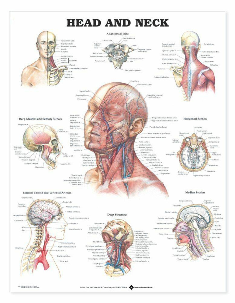
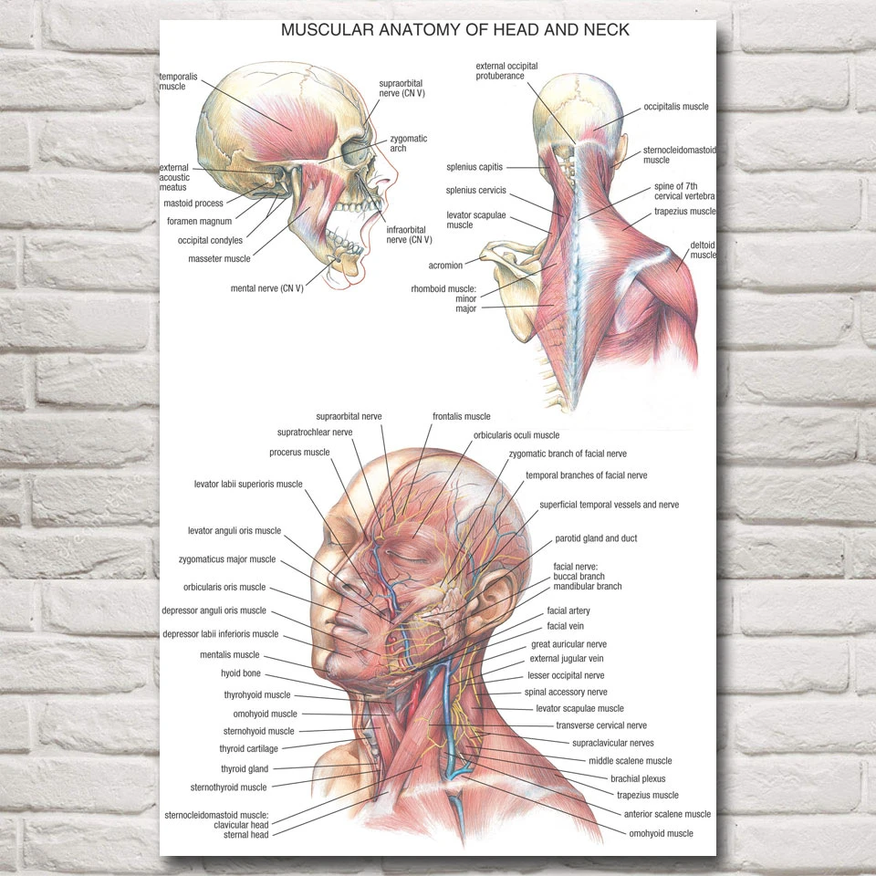
Head and Neck Chart 20x26 This 20" x 26" (51 x 66 cm) wall chart for examination rooms and classrooms shows the skull, brain, cervical spine, deep muscles and sensory nerves, internal carotid and vertebral arteries and deep structures of the face and shoulders illustrated by Peter Bachin. Horizontal and median sections are featured.

Head and neck laminated anatomy chart
Neck muscles work together with tendons and ligaments to support and move the neck and head. Tendons are connective tissue that attach muscle to bone, whereas ligaments attach bones to other bones.

Muscles of the head and neck
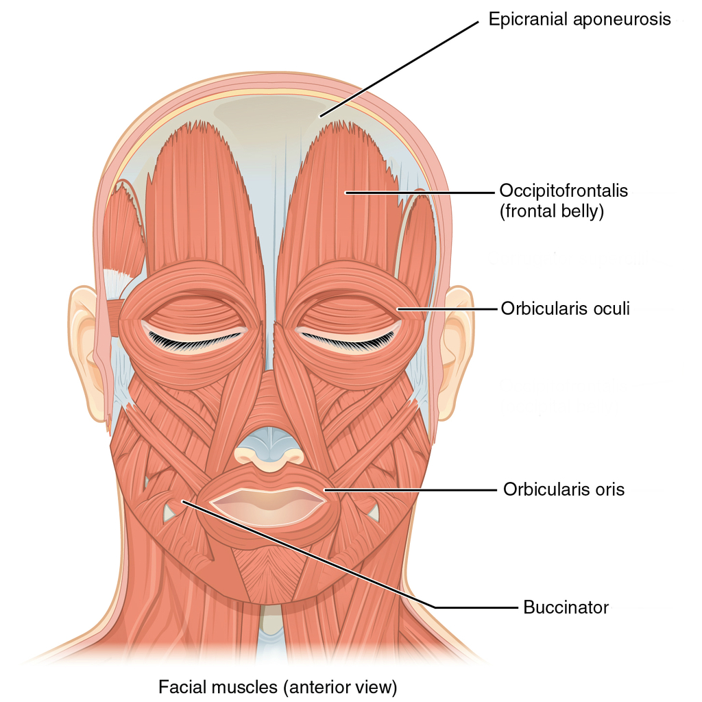
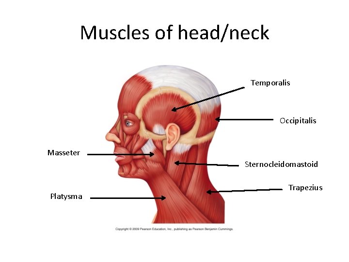
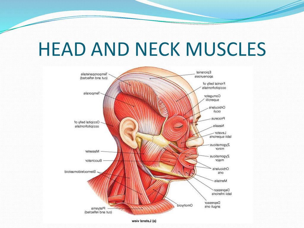
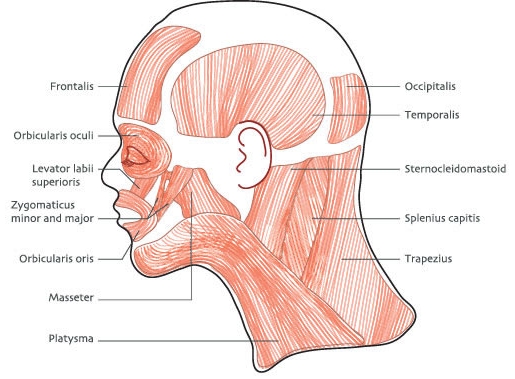
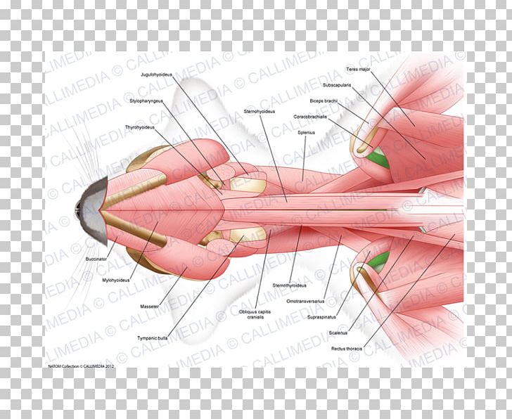
Head And Neck Muscles Diagram In this image, you will find cranial aponeurosis, temporalis, occipitalis, masseter, sternocleidomastoid, trapezius, platysma, orbicularis oris, buccinator, zygomaticus, orbicularis oculi, frontalis in Head and neck muscles diagram. Anatomy note Youtube Channel, Please Subscribe to support
Muscles of the head and neck - anatomy pictures and information
August 2, 2021 - Your neck muscles support your head and help you do a range of movements. They also assist with chewing, swallowing and breathing.

Anatomy of short neck muscles. short neck muscles with marked ...
Feb 23, 2014 - Muscles of The Head - Learn Human Anatomy

Head and neck muscles labeled anatomical diagram facial ...
Some of the axial muscles may seem to blur the boundaries because they cross over to the appendicular skeleton. The first grouping of the axial muscles you will review includes the muscles of the head and neck, then you will review the muscles of the vertebral column, and finally you will review ...

Anatomy of the neck and head stock photo - download image now ...
Blank Head and Neck Muscles Diagram. Why is it important to learn muscle anatomy? Muscle and anatomy are two words that are often heard when you are studying science. The human body consists of many muscles. If someone wants a healthy and good life, one must understand his body. How do you take care of a body if you don't know the anatomy?
Thumb muscle head and neck anatomy head and neck anatomy png ...
Instant anatomy is a specialised web site for you to learn all about human anatomy of the body with diagrams, podcasts and revision questions
Neck | anatomy | britannica
Feb 22, 2012 - This Pin was discovered by M@ryam Ahmadi. Discover (and save!) your own Pins on Pinterest
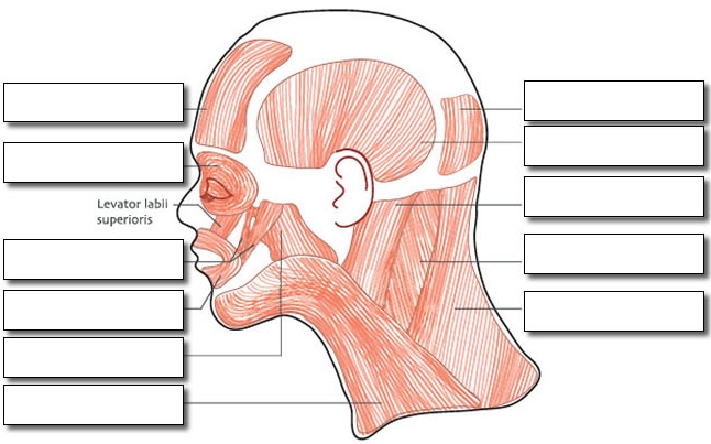
Label the muscles of the head
Several other muscles act on the head and neck. Below are three with a larger impact. Trapezius: The trapezius is the most superficial muscle of the back and forms a broad flat triangle. Attachments: The trapezius originates from the skull and spine of the upper back and neck. It attaches to the clavicle and scapula.

Fibromyalgia (fm) involved head-neck muscles. henry vandyke ...
0 Response to "38 head and neck muscle diagram"
Post a Comment