39 meiotic division beads diagram
meiotic division (prophase I and II, metaphase I and II, anaphase I and II, telophase I and II, and cytokinesis). Diagram the corresponding images for each stage in the sections titled "Trial 1 - Meiotic Division Beads Diagram". Be sure to indicate the number of chromosomes present in each cell for each phase. Disassemble the beads used ... Configure the chromosomes as they would appear in each of the stages of meiotic division (prophase I and II, metaphase I and II, anaphase I and II, telophase I and II, and cytokinesis). Diagram the corresponding images for each stage in the section titled "Trial 2 - Meiotic Division Beads Diagram".
Diagram the corresponding images for each stage in the sections titled "Trial 1 - Meiotic Division Beads Diagram". Be sure to indicate the number of chromosomes present in each cell for each phase. Disassemble the beads used in Trial 1. You will need to recycle these beads for a second meiosis trial in Steps 7 - 11.
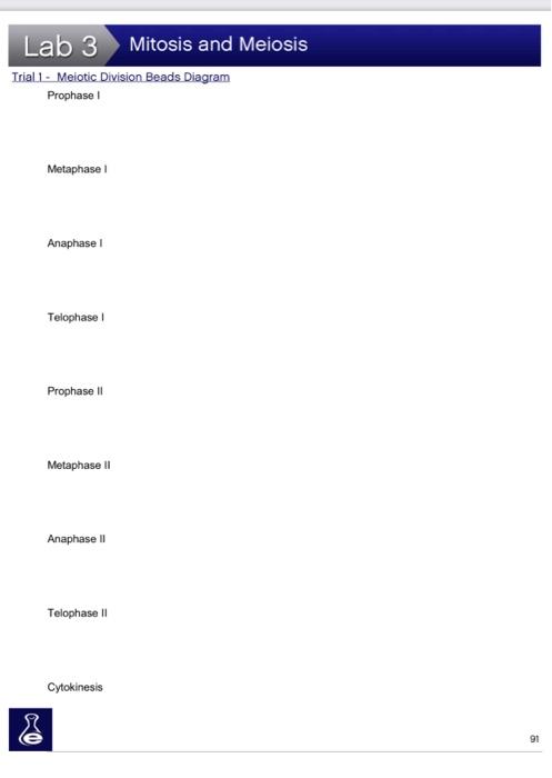
Meiotic division beads diagram
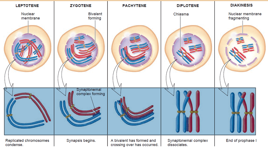
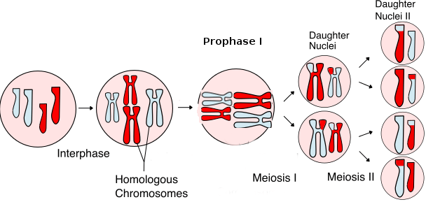
A chromosome is a long DNA molecule with part or all of the genetic material of an organism. Most eukaryotic chromosomes include packaging proteins called histones which, aided by chaperone proteins, bind to and condense the DNA molecule to maintain its integrity. These chromosomes display a complex three-dimensional structure, which plays a significant role in … Meiotic Division of Cell (With Diagram) In this article we will discuss about the meiotic division of a cell. The meiotic division includes two complete divisions of a diploid cell resulting into four haploid nuclei. The first meiotic division includes a long prophase in which the homologous chromosomes become closely associated to each other ... Diagram the corresponding images for each stage in the section titled "Trial 2 - Meiotic Division Beads Diagram". Be sure to indicate the number of chromosomes present in each cell for each phase. Also, indicate how the crossing over affected the genetic content in the gametes from Part1 versus Part 2. 95 Meiosis
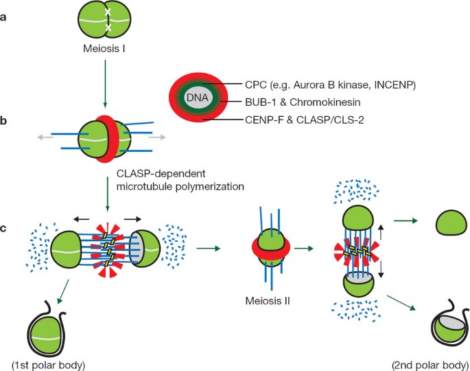
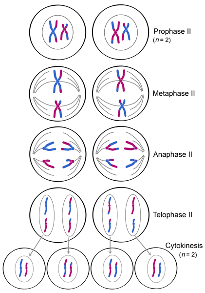
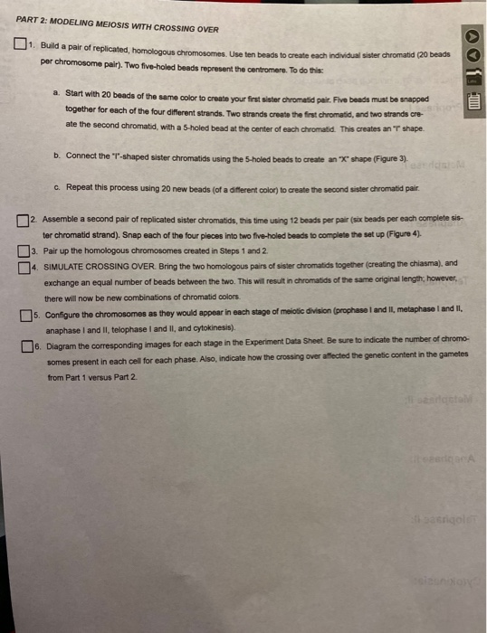
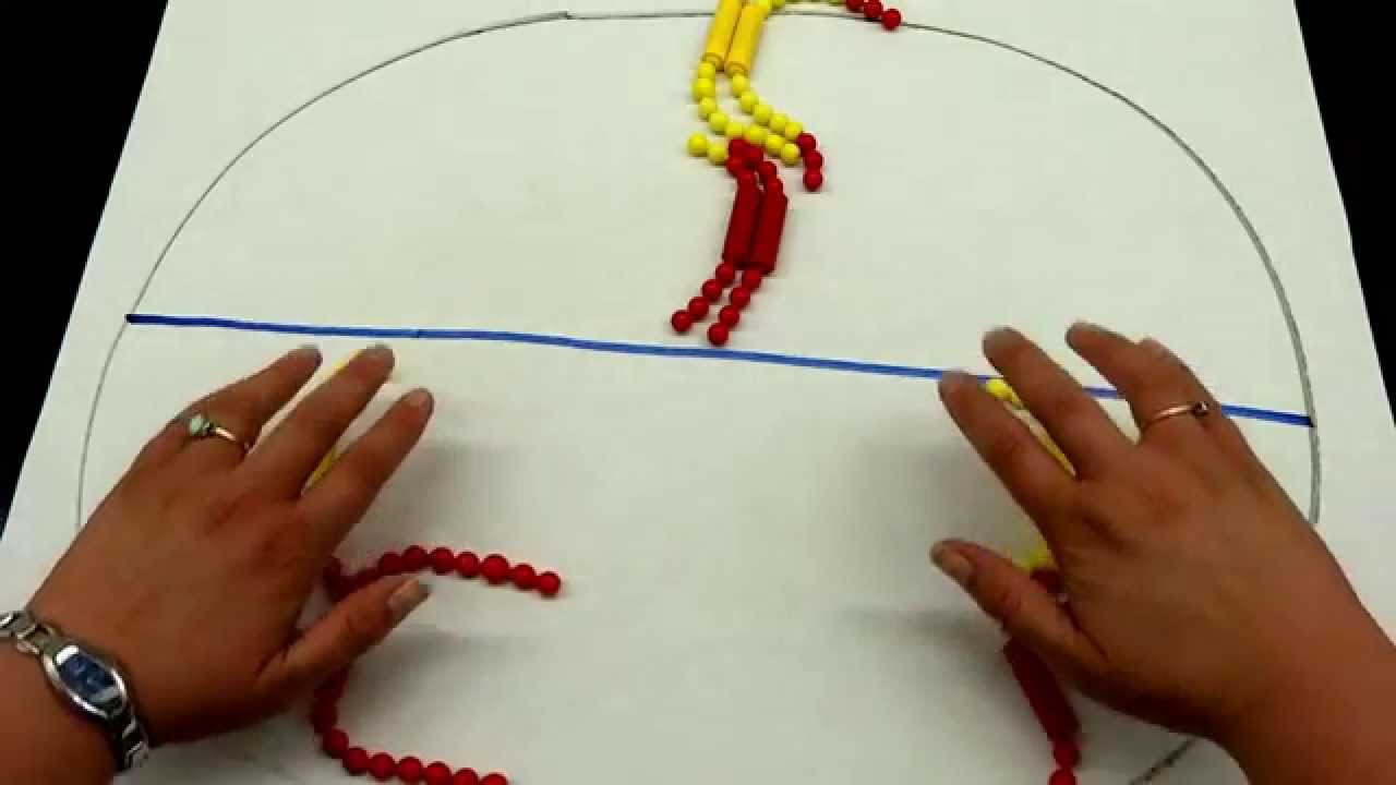
Meiotic division beads diagram. Part 1 - Meiotic Division Beads Diagram Prophase I Metaphase I Anaphase I Telophase I Prophase II Metaphase II Anaphase II 94 Meiosis Telophase II Cytokinesis Part 2: Modeling Meiosis with Crossing Over 7. Build a pair of replicated, homologous chromosomes. 10 beads should be used to create each individual Trial 1 - Meiotic Division Beads Diagram. Part 2: Modeling Meiosis with Crossing Over Part 2 - Meiotic Division Beads Diagram: Image of page 3. Info icon This preview has intentionally blurred sections.Lab 4: Meiosis and Vertebrate Reproduction LAB SYNOPSIS: • Meiosis will be modeled using pop-beads. • The genetic diversity of gametes will ... 27.1.2021 · Binding of METTL3 to chromatin is enriched over IAP family endogenous retroviral elements in mouse embryonic stem cells, helping to ensure the integrity of … The second meiotic division is where sister (duplicated) chromatids separate. It resembles mitosis of a haploid cell. At the start of the second division, each cell contains 1N chromosomes, each consisting of a pair of sister chromatids joined at the centromere. Here is a simplified diagram illustrating the overall process and products of meiosis:
Who are the experts? Experts are tested by Chegg as specialists in their subject area. We review their content and use your feedback to keep the quality high. 100% (2 ratings) Meiosis comprises of two phases. In phase one the div …. View the full answer. Transcribed image text: Trial 2 - Meiotic Division Beads Diagram. Your Full Name: 102/103 Lab 5: Meiosis INSTRUCTIONS: and submit it via the Assignments Folder by the date listed in the Course Schedule (under Syllabus). The male gonad is the testis (pl, testes).. The initial difference in male and female gonad development are dependent on testis-determining factor (TDF) the protein product of the Y chromosome SRY gene. Recent studies have indicated that additional factors may also be required for full differentiation. Experiment 1: Following Chromosomal DNA Movement through Meiosis Data Tables and Post-Lab Assessment Trial 1 - Meiotic Division Beads Diagram: Biol 103 papers , exams and assignments and many more for students. At Maryland homework we offer assignments and exams from students just like you who have got A grades on these papers.
second meiotic division (meiosis II) that they finally are separated and distributed into separate ... In this diagram the chromosomes are shown as if they were visible, ... which clearly defined beads of local coiling (chromomeres) can be seen. Part 1 - Meiotic Division Beads Diagram Prophase I Metaphase I Anaphase I Telophase I Prophase II Metaphase II Anaphase II Telophase II Cytokinesis Part 2: Modeling Meiosis with Crossing Over Build a pair of replicated, homologous chromosomes. 10 beads should be used to create each individual sister chromatid (20 beads per chromosome pair). Diagram the corresponding images for each stage in the section titled "Trial 2 - Meiotic Division Beads Diagram". Be sure to indicate the number of chromosomes present in each cell for each phase. Also, indicate how the crossing over affected the genetic content in the gametes from Part1 versus Part 2. Part 2 - Meiotic Division Beads ... Diagram the corresponding images for each stage in the sections titled "Trial 1 - Meiotic Division Beads Diagram". Be sure to indicate the number of chromosomes present in each cell for each phase. Disassemble the beads used in Part 1. You will need to recycle these beads for a second meiosis trial in Steps 7 - 12. Trial 1 - Meiotic Beads Diagram:
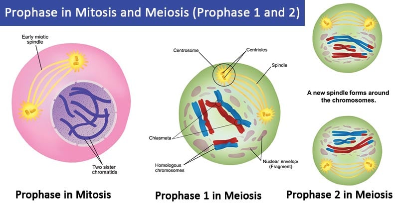
Prophase in mitosis and meiosis (Prophase 1 and 2)
Your Full Name: 102/103. Lab 5: Meiosis. INSTRUCTIONS: and submit it via the Assignments Folder by the date listed in the Course. Schedule (under Syllabus). To conduct your laboratory exercises, use the Laboratory Manual located under. Course Content.
ACTIVITY: Modeling Meiosis with Pop Beads
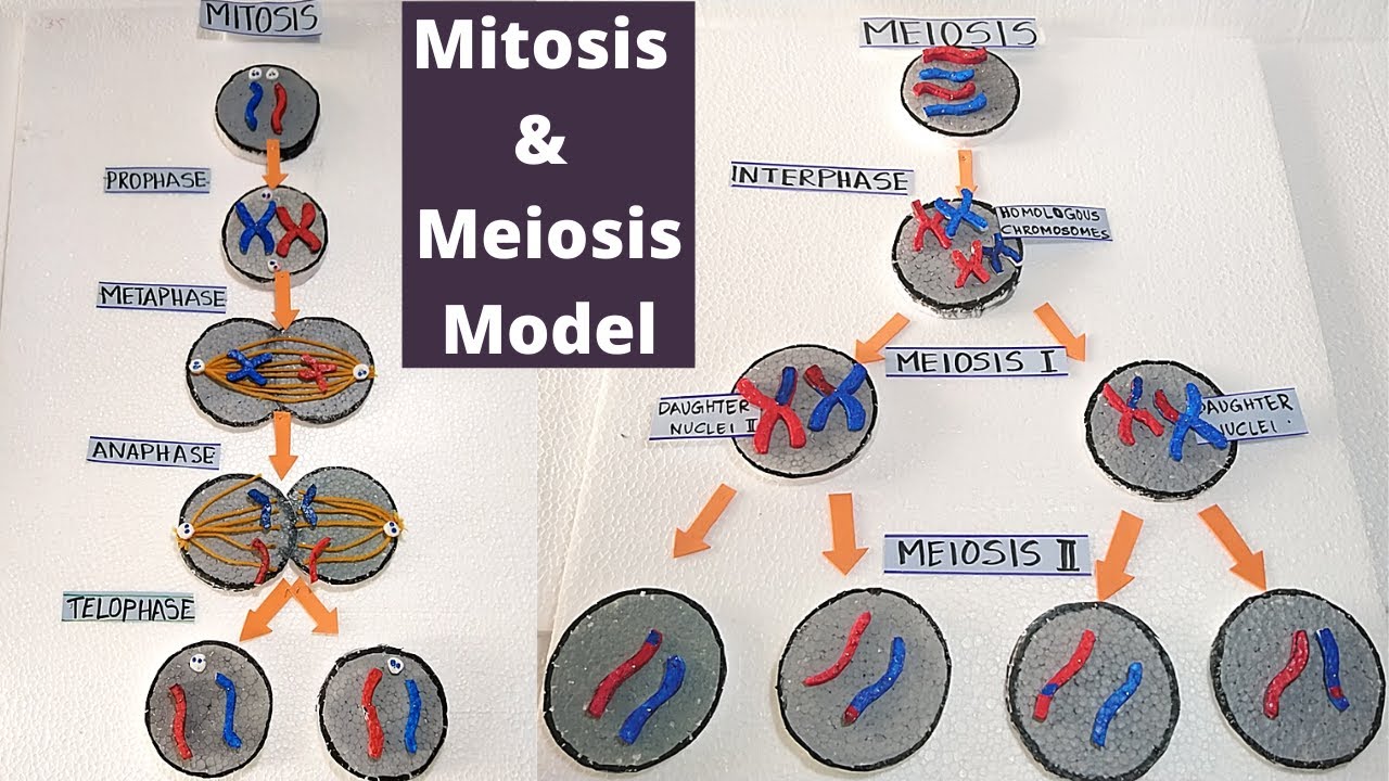
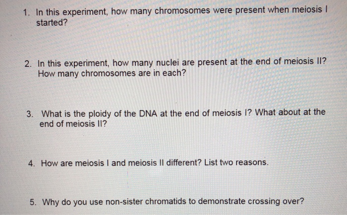
11. Diagram what would happen if sexual reproduction took place for four generations using diploid (2n) cells. Since mitosis produces daughter cells with the diploid number of chromatids, it cannot be used for sperm or egg production. If a 2N (diploid) sperm united with a 2N egg, the results would be disastrous (4N). The fertilized egg would have twice the amount of genetic material it must have.
Prophase I - Definition, Stages and Quiz | Biology Dictionary
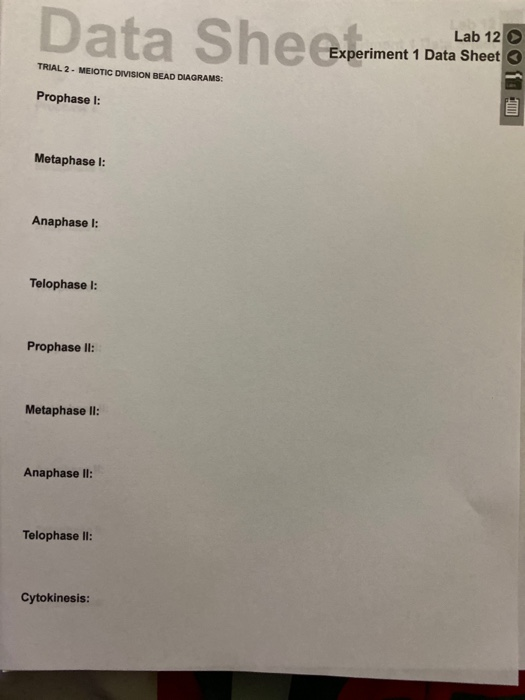
Transcribed image text: Data She experi Lab 12 > Experiment 1 Data Sheet dels TRIAL 2 - MEIOTIC DIVISION BEAD DIAGRAMS: Prophase : LE Metaphase 1: Anaphase 1: Telophase 1: Prophase II: Metaphase II: Anaphase II: Telophase II: Cytokinesis: Data SheeExperia Lab 12 Experiment 1 Data Sheet TRIAL 2 - MEIOTIC DIVISION BEAD DIAGRAMS: Prophase 1 ...
Mitosis (article) | Cellular division | Khan Academy
stages of meiotic division (prophase I and II, metaphase I and II, anaphase I and II, telophase I and II, and cytokinesis). 12. Diagram the corresponding images for each stage in the section titled "Trial 2 Meiotic Division Beads Diagram". Be sure to indicate the number of chromosomes present in each cell for each phase. Also, indicate how ...
Diffusion-mediated HEI10 coarsening can explain meiotic ...
Part 1 - Meiotic Division Beads Diagram Prophase I Metaphase I Anaphase I Telophase I Prophase II Metaphase II Anaphase II Telophase II Cytokinesis Part 2: Modeling Meiosis with Crossing Over Build a pair of replicated, homologous chromosomes. 10 beads should be used to create each individual sister chromatid (20 beads per chromosome pair).

Regulated Proteolysis of MutSγ Controls Meiotic Crossing Over ...
stages of meiotic division (prophase I and II, metaphase I and II, anaphase I and II, telophase I and II, and cytokinesis). 12. Diagram the corresponding images for each stage in the section titled "Trial 2 Meiotic Division Beads Diagram". Be sure to indicate the number of chromosomes present in each cell for each phase. Also, indicate how ...
Phosphoregulation of HORMA domain protein HIM-3 promotes ...
Griffiths - Introduction to Genetic Analysis 9th Edition
Solved Data She experi Lab 12 > Experiment 1 Data Sheet dels ...
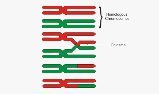
The meiotic board used includes six circles: one on top; two in the middle and four at the bottom. These circles represent the chromosomal make up of the seven nuclei involved in one meiotic event. The top circle represents the karyotype of mother nucleus, the center two of the two daughter nuclei, and the bottom four of the granddaughter nuclei.

IB_1108_L12_Meiosis.docx - Meiosis PRE-LAB QUESTIONS 1 ...
11.11.2021 · The cereal endosperm is a major determinant of seed size and shape. Here the authors show that a lncRNA, MISSEN, is expressed from the maternally derived allele in rice seeds and regulates a ...

IB_1108_L12_Meiosis.docx - Meiosis EXPERIMENT 1 FOLLOWING ...
Diagram the corresponding images for each stage in the section titled "Trial 2 - Meiotic Division Beads Diagram". Be sure to indicate the number of chromosomes present in each cell for each phase. Also, indicate how the crossing over affected the genetic content in the gametes from Part1 versus Part 2.
Prophase in mitosis and meiosis (Prophase 1 and 2)
View L12_Meiosis.docx from BSC1005L 1005 at Broward College. Meiosis EXPERIMENT 1: FOLLOWING CHROMOSOMAL DNA MOVEMENT THROUGH MEIOSIS Trial 1 - Meiotic Division Beads Diagram Prophase I Metaphase
ACTIVITY: Modeling Meiosis with Pop Beads
In cell biology, the spindle apparatus (or mitotic spindle) refers to the cytoskeletal structure of eukaryotic cells that forms during cell division to separate sister chromatids between daughter cells.It is referred to as the mitotic spindle during mitosis, a process that produces genetically identical daughter cells, or the meiotic spindle during meiosis, a process that produces …
Meiotic kinetochores get pushed aside by a CLS act | Nature ...
Part 1 - Meiotic Division Beads Diagram Prophase I Metaphase I Anaphase I Telophase I Prophase II Metaphase II Anaphase II Telophase II Cytokinesis Part 2: Modeling Meiosis with Crossing Over Build a pair of replicated, homologous chromosomes. 10 beads should be used to create each individual sister chromatid (20 beads per chromosome pair).
Life Sciences Cyberbridge
Trial 1 - Meiotic Division Without Crossing Over Beads Diagram: Take pictures of your beads for each phase of meiosis I and II without crossing over. Include notes with your name, date and meiotic stage on index cards in the pictures. Please use the lowest resolution possible so that your file does not become too large to submit.
Solved] Experiment 1: Following Chromosomal DNA Movement ...
Diagram the corresponding images for each stage in the sections titled "Trial 1 - Meiotic Division Beads Diagram". Be sure to indicate the number of chromosomes present in each cell for each phase. Disassemble the beads used in Trial 1. You will need to recycle these beads for a second meiosis trial in Steps 7 - 11.
ACTIVITY: Modeling Meiosis with Pop Beads
102/103. Lab 5: Meiosis. INSTRUCTIONS: and submit it via the Assignments Folder by the date listed in the Course. Schedule (under Syllabus). To conduct your laboratory exercises, use the Laboratory Manual located under. Course Content. Read the introduction and the directions for each exercise/experiment.
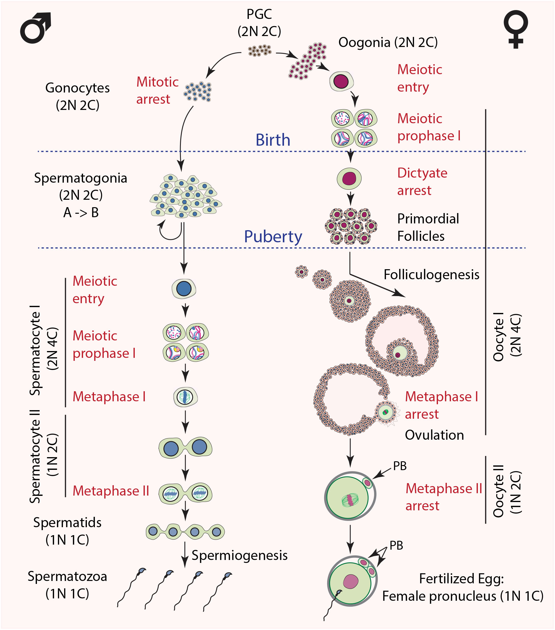
Meiosis: the chromosomal foundation of reproduction
Diagram the corresponding images for each stage in the section titled "Trial 2 - Meiotic Division Beads Diagram". Be sure to indicate the number of chromosomes present in each cell for each phase. Also, indicate how the crossing over affected the genetic content in the gametes from Part1 versus Part 2.
SUMO is a pervasive regulator of meiosis | eLife
Budding yeast continues to propel our understanding of regulated cell division: critical studies in the structure and regulation of signal transduction pathways (Mok et al. 2011), subcellular organization (Taddei and Gasser 2012), movement of key regulators (D’Amours and Amon 2004), checkpoint regulation [of DNA repair, spindle function, cellular growth, nutrient response, and …

Experiment 3 ap1 lab3.docx - EXPERIMENT 3 FOLLOWING ...
16.1.2022 · To address which mitochondria-related nuclear differentially expressed genes (DEGs) and related pathways are altered during human oocyte maturation, single-cell analysis was performed in three oocyte states: in vivo matured (M-IVO), in vitro matured (M-IVT), and failed to mature in vitro (IM-IVT). There were 691 DEGs and 16 mitochondria-related DEGs in the …
ROLE OF 14-3-3 ETA AND EPSILON IN GAMETOGENESIS
In experiment of Following chromosomal DNA movement through meiosis, what is the trial 1 and trial 2 meiotic division beads diagram for prophase l, metaphase l, anapahse l, telophase l, prophase ll, metaphase ll, anaphase ll, telophase ll, and cytokinesis? Start your trial now! First week only $4.99! arrow_forward.
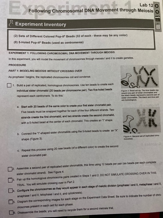
Solved Data She experi Lab 12 > Experiment 1 Data Sheet dels ...
Diagram the corresponding images for each stage in the section titled "Trial 2 - Meiotic Division Beads Diagram". Be sure to indicate the number of chromosomes present in each cell for each phase. Also, indicate how the crossing over affected the genetic content in the gametes from Part1 versus Part 2. 95 Meiosis

The Proteomic Landscape of Centromeric Chromatin Reveals an ...
Meiotic Division of Cell (With Diagram) In this article we will discuss about the meiotic division of a cell. The meiotic division includes two complete divisions of a diploid cell resulting into four haploid nuclei. The first meiotic division includes a long prophase in which the homologous chromosomes become closely associated to each other ...

Simulating meiotic homolog pairing specificity. (A) Diagram ...
A chromosome is a long DNA molecule with part or all of the genetic material of an organism. Most eukaryotic chromosomes include packaging proteins called histones which, aided by chaperone proteins, bind to and condense the DNA molecule to maintain its integrity. These chromosomes display a complex three-dimensional structure, which plays a significant role in …

Cell division: mitosis and meiosis | Biological Principles
IJMS | Free Full-Text | Putative Origins of Cell-Free DNA in ...

Mitosis and Meiosis Simulation
Frontiers | An Optimized and Versatile Counter-Flow ...
Solved Data She experi Lab 12 > Experiment 1 Data Sheet dels ...
Mitosis & Meiosis Cell Division Model | mitosis and Meiosis model | How to make Cell division Model
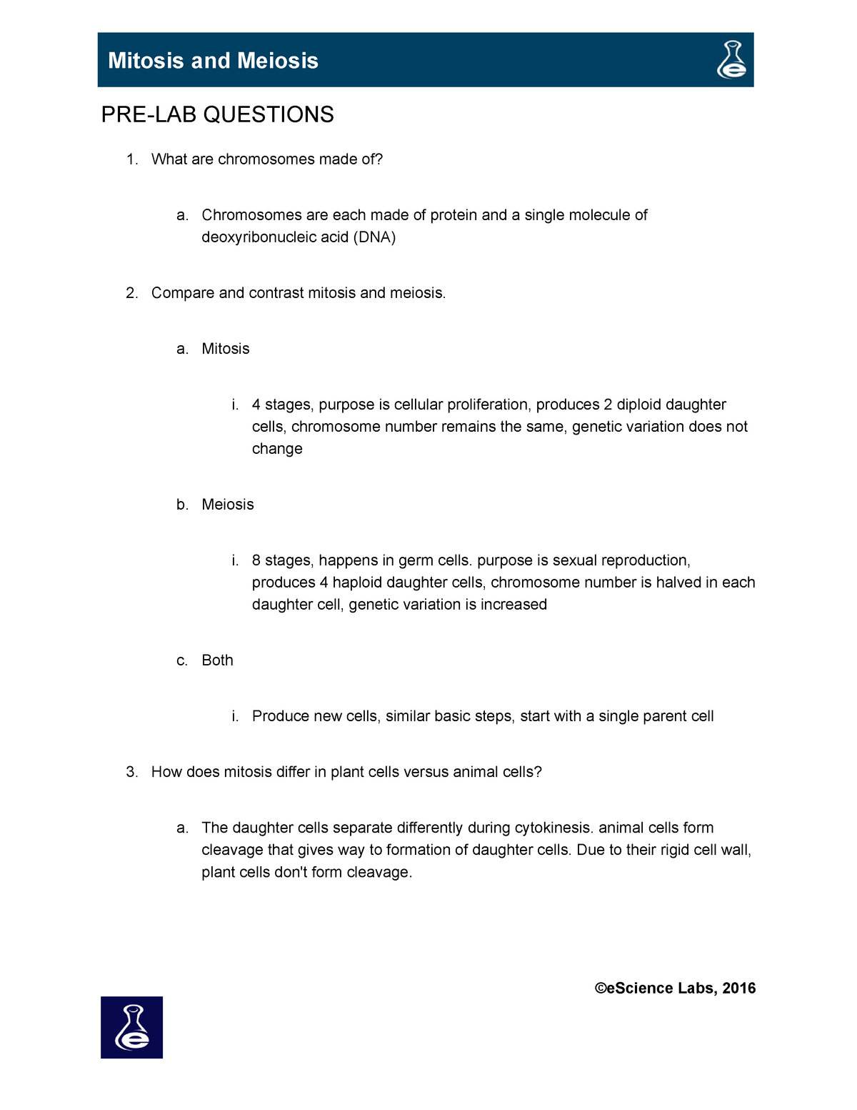
Mitosis and Meiosis Lab - PRE-LAB QUESTIONS What are ...
Solved Lab 3 3 Mitosis and Meiosis Experiment 3: Following ...
Solved AA- A - 2017 - A-D-A- ÐавьÑct AABBC ÐавьÑÑ ÐавьÑ. 1 ...
ragraph Trial 1- Meiotic Beads Diagram: Prophase l | Chegg.com

Meiosis_Lab-converted.pdf - EXPERIMENT 1 FOLLOWING ...
Xkid chromokinesin is required for the meiosis I to meiosis ...
Biochemical and functional characterization of a meiosis ...

Cell Cycle Division Mitosis Beads Diagram - Wiring Site Resource
Lab 10: Part 1 - Meiosis bead demonstration
Cell division: mitosis and meiosis | Biological Principles
0 Response to "39 meiotic division beads diagram"
Post a Comment