40 inferior vena cava diagram
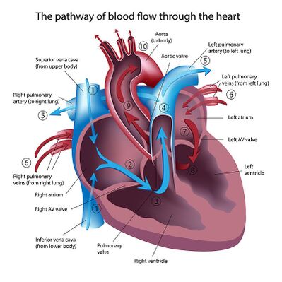
Inferior Vena Cava (IVC) - Geeky Medics Overview of the inferior vena cava. The IVC is formed by the union of the right and left common iliac veins.It conveys systemic venous blood from the lower limbs and pelvis, the undersurface of the diaphragm and parts of the abdominal wall.The IVC does not drain blood from the gut.. Course of the IVC. The IVC begins in the abdomen at L5 and ends in the thorax at T8, where it enters the ... diagram of how blood flows through the heart - Lisbdnet.com 38 Box Diagram, Labels of Heart, and Blood Flow through Heart; ... What is the order of blood flow through the heart assume we are starting at the superior and inferior vena cava? The blood enters the heart from the body through the superior vena cava and the inferior vena cava. Then the blood enters the right atrium chamber of the heart.
Echocardiographic Evaluation of Pericardial Effusion and ... 24.04.2017 · Typical image of inferior vena cava (IVC) plethora (left) in a patient with significant pericardial effusion. Collapse of IVC during inspiration is less than 50% (right). (v) Septal “bounce” An inspiratory “bounce” of the interventricular septum toward the LV is a common, but not specific finding in cardiac tamponade (Figure (Figure12). 12). An M-mode through the parasternal long …
Inferior vena cava diagram
Brachial Artery Location, Anatomy, and Function - Healthline 06.03.2019 · The inferior vena cava is also referred to as the posterior vena cava. The inferior vena cava is a large vein that carries deoxygenated blood from the… READ MORE. Arcuate artery of the foot ... Inferior Vena Cava: Anatomy, Function, and Significance The inferior vena cava (also known as IVC or the posterior vena cava) is a large vein that carries blood from the torso and lower body to the right side of the heart. From there the blood is pumped to the lungs to get oxygen before going to the left side of the heart to be pumped back out to the body. The IVC gets its name from its structure ... Schematic diagram showing embryogenesis of inferior vena ... Schematic diagram showing embryogenesis of inferior vena cava and renal veins. A. Three pairs of veins (posterior cardinal → subcardinal → supracardinal veins) appear in succession with ...
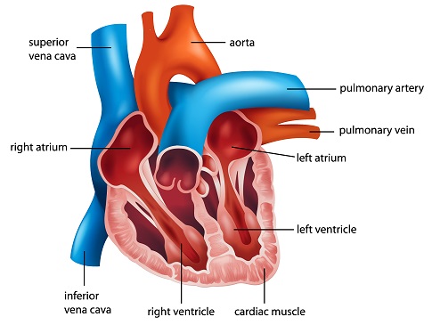
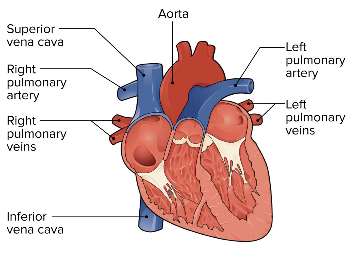
Inferior vena cava diagram. commons.wikimedia.org › wiki › File:Diagram_of_theFile:Diagram of the human heart (cropped).svg - Wikimedia Feb 28, 2022 · English: Diagram of the human heart. 1. Superior vena cava 2. 4. Mitral valve 5. Aortic valve 6. Left ventricle 7. Right ventricle 8. Left atrium 9. Right atrium 10. Aorta 11. Pulmonary valve 12. Tricuspid valve. 13. Inferior vena cava Diagram of Blood Flow Through the Heart - Bodytomy The right and left side or chambers of the heart work in tandem with each other. Blood from all over the body will reach the heart through pulmonary veins; inferior vena cava and the superior vena cava. The deoxygenated blood will first enter the right atrium and simultaneously the left atrium receives oxygenated blood from the lungs. PDF Anatomy of Heart Labeled and Unlabeled Images Superior vena cava Ascending aorta Right pulmonary artery Pulmonary trunk Right pulmonary veins Auricle of right atrium Right coronary artery (in atrioventricular sulcus) Right ventricle Inferior vena cava (a) Anterior view of the external heart C' 2019 Pearson Education. Aort'c arch Ligamentum arteriosum Left pulmonary artery Left pulmonary ve ns In the above given diagram which blood vessel represents ... The superior vena cava carry deoxygenated blood from the head, chest, upper limbs to the right auricle. The inferior vena cava carry deoxygenated blood from the abdomen, lower limbs to the right auricle. So, the correct answer is option B.
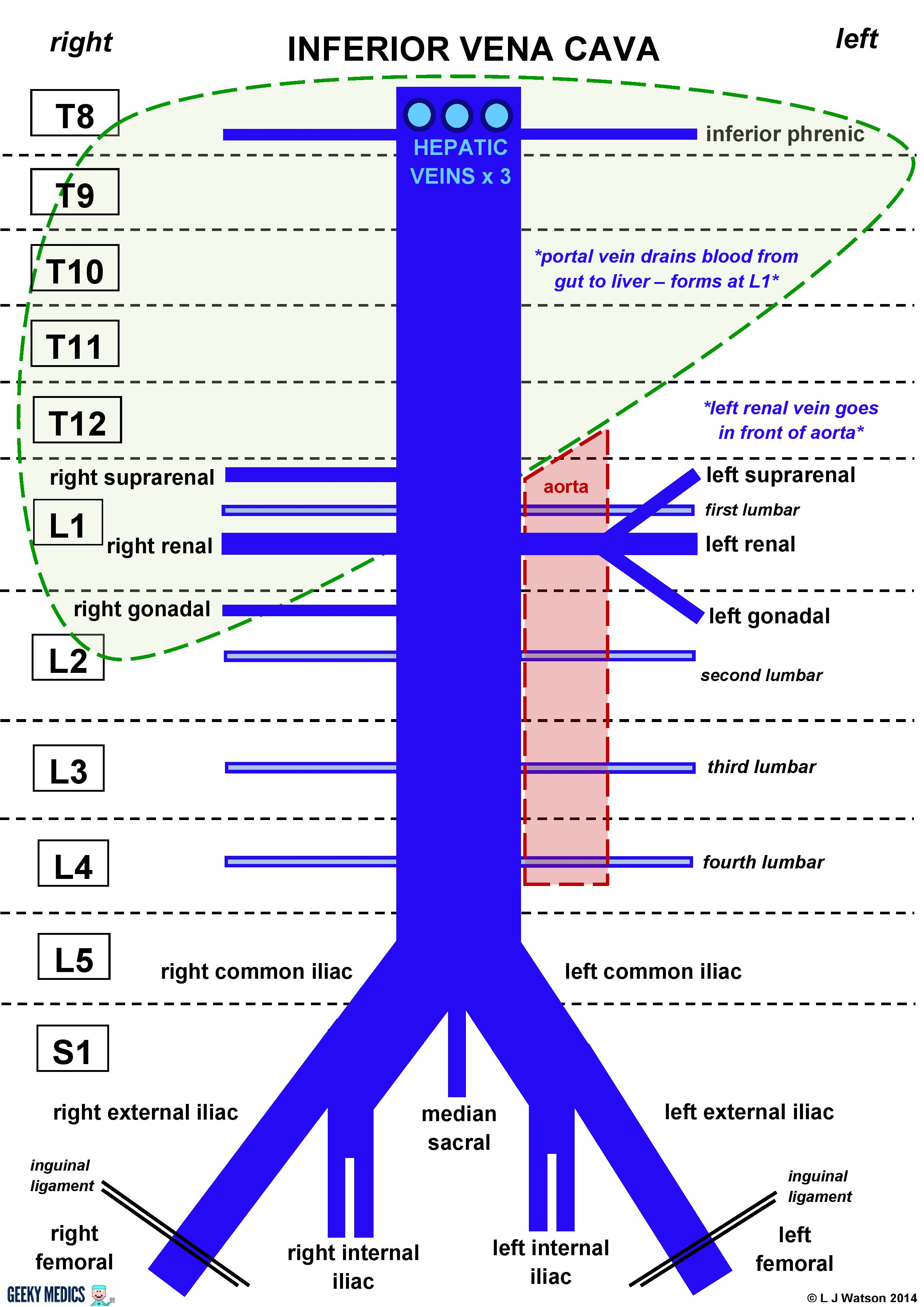
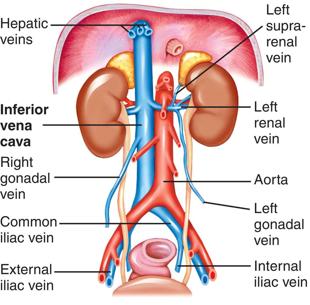
Circulatory system Facts for Kids - Kiddle 14.12.2021 · The inferior vena cava takes blood from the lower part of the body to the right side of the heart. (In medicine, inferior means below.) ... Above is a diagram of an open circulatory system. An open circulatory system is made up of a heart, vessels, and hemolymph. This diagram shows how the hemolymph, fluid present in most invertebrates that is equivalent to … en.wikipedia.org › wiki › Umbilical_veinUmbilical vein - Wikipedia Fetal circulation. The unpaired umbilical vein carries oxygen and nutrient rich blood derived from fetal-maternal blood exchange at the chorionic villi.More than two-thirds of fetal hepatic circulation is via the main portal vein, while the remainder is shunted from the left portal vein via the ductus venosus to the inferior vena cava, eventually being delivered to the fetal right atrium. Renal vein - Wikipedia Because the inferior vena cava is on the right half of the body, the left renal vein is generally the longer of the two. Because the inferior vena cava is not laterally symmetrical, the left renal vein often receives the following veins: left inferior phrenic vein; left suprarenal vein; left gonadal vein (left testicular vein in males, left ovarian vein in females) left 2nd lumbar vein; This ... Urinary System - 2nd Period Group 7 Tennis - Google Search Inferior Vena Cava- Takes deoxygenated blood from the kidney back to the heart. Renal Artery- Takes oxygenated blood from the Abdominal Aorta to the kidneys. Renal Vain- Takes deoxygenated blood from the kidney to the Inferior Vena Cava. Kidneys- Filter waste and water together into urine. Ureter- tubes that take the urine to the bladder.
Common Iliac Vein Anatomy, Function, and Diagram | Body Maps The inferior vena cava is also referred to as the posterior vena cava. The inferior vena cava is a large vein that carries deoxygenated blood from the… READ MORE Migrated Inferior Vena Cava (IVC) Filter Strut: A Rare ... BACKGROUND Inferior vena cava (IVC) filters are indicated for patients with recurrent venous thrombosis despite proper anticoagulation or whenever anticoagulation is contraindicated. IVC filter deployment is an invasive procedure with various complications. One example is IVC filter limb fracture an … Heart Model Diagram | Quizlet Superior Vena Cava... Inferior Vena Cava... Pulmonary Veins... Coronary Sulcus... Posterior interventricular sulcus... Other sets by this creator. Coronary Vessels. 4 terms. Zack_Tolley PLUS. Structures of Dissected Heart. 12 terms. Zack_Tolley PLUS. Inferior Vena Cava - Anatomy Pictures and Information Inferior Vena Cava. The inferior vena cava is the largest vein in the human body. It collects blood from veins serving the tissues inferior to the heart and returns this blood to the right atrium of the heart. Although the vena cava is very large in diameter, its walls are incredibly thin due to the low pressure exerted by venous blood.
Inferior vena cava: Anatomy and function | Kenhub
Fetal Circulation Diagram | Fetal Blood Flow & Circulatory ... The inferior vena cava takes blood to the right atrium of the heart Through a series of shunts and openings, the blood flows through the heart bypassing the lungs.

Medical Definition of Inferior vena cava
PDF Name: Heart Anatomy Coloring - Weebly inferior vena cava. The superior vena cava carries oxygen-poor blood from the head and upper limbs. The inferior vena cava carries oxygen-poor blood from the abdomen and lower limbs. The deoxygenated blood enters the right atrium of the heart. Then the blood passes through the tricuspid valve to the right ventricle.
Inferior Vena Cava (IVC) Filter Placement - Atlanta Vascular ...
Circulatory System: Blood Flow Pathway Through the Heart ... Superior Vena Cava & Inferior Vena Cava. The vena cava is the largest vein in the body that delivers oxygen-poor or deoxygenated blood to the right atrium of the heart. The superior vena cava comes from the upper part of the body, including the brain and arms, while the inferior vena cava comes from the abdominal area and legs.

Inferior Vena Cava Clip Art - Royalty Free - GoGraph
Inferior vena cava diagram | scienceforyou Inferior Vena Cava Diagram. There are several key points to take away from this diagram. Although the vena cava is very large in diameter its walls are incredibly thin due to the low pressure exerted by venous blood. In this image you will find hepatic veins inferior phrenic vein portal vein left renal vein left suprarenal vein left gonadal ...
Vena Cava - Physiopedia
Observe the diagram and fill in the box with the ... Observe the diagram and fill in the box with the appropriate choice. A. Inferior vena cava. B. Superior vena cava. C. Pulmonary vein. D. Renal artery. Solution. The correct option is B. Superior vena cava. The superior vena cava carries deoxygenated blood from the upper half of the body to the heart. It drains the blood into the right auricle.

Double Inferior Vena Cava disease: Malacards - Research ...
Atrial Septal Defect | Congenital Heart Disease - Cove ... Inferior Vena Caval Variations of ASD Atrial Septal Defects are divided into three different types on the basis of the position of the hole (or holes) in the atrial septum. The first type of ASD is known as ostium primum defect, or simply, primum (number 1 in the diagram).

Superior Vena Cava Syndrome: Symptoms, Treatments, Causes
PDF The Cardiovascular System - Pearson the inferior mediastinum (me″de-as-ti′num), the medial section of the thoracic cavity, the heart is (a) Superior vena cava Left lung Aorta Parietal pleura (cut) Pericardium (cut) Pulmonary trunk Diaphragm Apex of heart Figure 11.1 Location of the heart within the thorax. (a) Relationship of the

inferior vena cava | anatomy | Britannica
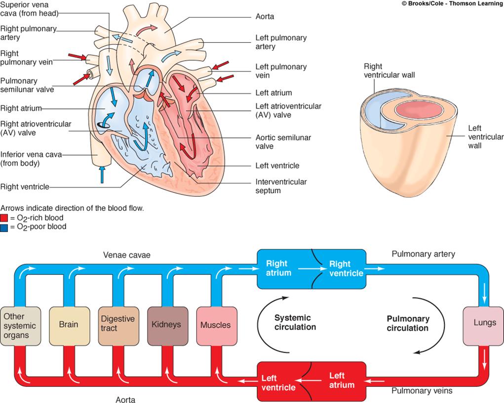
The Heart and Circulation of Blood - LSA All of the blood from the body is eventually collected into the two largest veins: the superior vena cava, which receives blood from the upper body, and the inferior vena cava, which receives blood from the lower body region. Both venae cavae empty the blood into the right atrium of the heart. From here the blood begins its journey through the pulmonary cycle. From the right …

Human Heart Circulatory System Diagram Chart Medical Educational Science Class Anatomy Corazon Veins Arteries Labels Cool Wall Decor Art Print Poster ...
Heart Blood Flow | Simple Anatomy Diagram, Cardiac ... Step 1 involves the superior vena cava (SVC) and inferior vena cava (IVC). They are the main blood vessels that carry the deoxygenated venous blood from the rest of the body to the right side of the heart, specifically the right atrium. The superior vena cava is located superiorly, and it carries the deoxygenated venous blood from the upper ...

Pin on Atlas anatomy
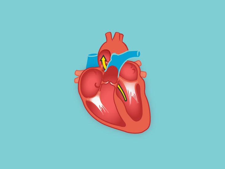
Flow Of Deoxygenated Blood Through The Heart - Etfatehran.net The pathway of blood flow through the heart begins as blood comes from the body and enters the heart through the superior and inferior vena cava indicated by the yellow star in the diagram below. Superior and inferior vena cavae and the coronary sinus 2. The right atrium receives deoxygenated blood through the superior and inferior vena cavas ...

Inferior vena cava | Radiology Reference Article ...
Inferior Vena Cava Syndrome - StatPearls - NCBI Bookshelf Inferior vena cava syndrome (IVCS) is a sequence of signs and symptoms that refers to obstruction or compression of the inferior vena cava (IVC). The pathophysiology of IVCS is similar to superior vena cava syndrome (SVCS) because of the presence of an underlying process that inhibits venous return to the right atrium. IVCS is not a primary diagnosis because it is often caused by other ...
Inferior Vena Cava (IVC) | Geeky Medics
› science › stomachstomach | Definition, Function, Structure, Diagram, & Facts ... Stomach, saclike expansion of the digestive system, between the esophagus and the small intestine; it is located in the anterior portion of the abdominal cavity in most vertebrates. The stomach serves as a temporary receptacle for the storage and mechanical distribution of food before it is passed into the intestine.

Superior & Inferior Vena Cava Function & Location | What ...
Heart Anatomy: Labeled Diagram, Structures, Blood Flow ... 24.02.2021 · Superior/Inferior Vena Cava. Now that we understand the blood flow to and from the heart, we can discuss the final structures. The first 2 structures are responsible for carrying deoxygenated blood from the body to the right side of the heart (right atrium). They are known as the superior vena cava and inferior vena cava.

Lower Branches of the Aorta & inferior vena cava Diagram ...
Inferior Vena Cava Function, Anatomy & Definition | Body Maps The inferior vena cava runs posterior, or behind, the abdominal cavity. This vein also runs alongside the right vertebral column of the spine. The inferior vena cava is the result of two major leg...

Inferior vena cava filter Superior vena cava Venae cavae Vein ...
Right Atrium - Location, Structure, Function, Diagram The blood enters the right atrium through inferior and superior vena cava. Location. Contents. Location; Structure; Function; Significance ; The right atrium is located in the upper right corner of the heart above the right ventricle. There is a tricuspid valve present between the right atrium and right ventricle. Structure. Diagram of Right Atrium. The right atrium muscular walls are …
Inferior vena cava: Anatomy and function | Kenhub
Blood vessels of abdomen and pelvis : Anatomy ... - Kenhub The inferior vena cava (IVC) is the headmaster of the veins department. It collects all the blood from the abdomen, pelvis and lower limbs and carries it to the right atrium of the heart . The IVC is formed by merging of the left and right common iliac veins at the L5 vertebral level, just in front of the aortic bifurcation.

371 Inferior Vena Cava High Res Illustrations - Getty Images
Inferior vena cava: Anatomy and function - Kenhub The inferior vena cava (IVC) is the largest vein of the human body. It is located at the posterior abdominal wall on the right side of the aorta. The IVC's function is to carry the venous blood from the lower limbs and abdominopelvic region to the heart.. The inferior vena cava anatomy is essential due to the vein's great drainage area, which also makes it a hot topic for anatomy exams.
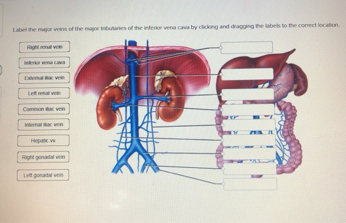
Solved Label the major veins of the major tributaries of the ...
› diaphragmDiaphragm: Definition, Location, Anatomy, Function, Diagram Sep 09, 2017 · Caval opening (vena caval hiatus) at the 8 th vertebral level: Allows the inferior vena cava [9] and the right phrenic nerve branches [11] to pass through. Esophageal opening (esophageal hiatus) at the 10th vertebra level: Transmits the esophagus, vagus nerves, and the small esophageal arteries [12] .
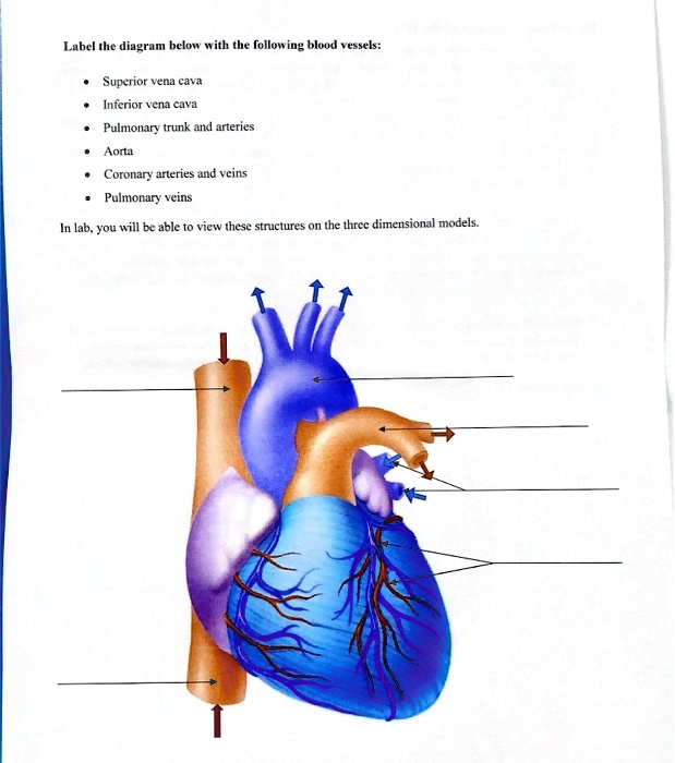
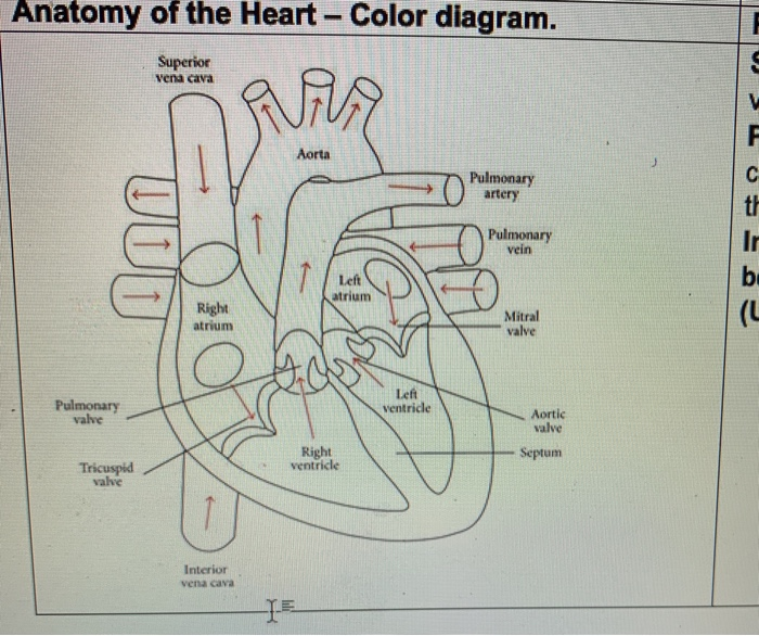
SOLVED:Label the diagram below #ith tbe following bloud ...
› treatments › staged-reconstructionStaged Reconstruction Heart Surgery - Children's Hospital of ... During this surgery the inferior vena cava, a large vein that carries deoxygenated blood from the lower body into the heart, is disconnected from the heart and attached to the pulmonary artery. After this operation, all of the deoxygenated blood from the body goes to the lungs without passing through the heart.

Difference Between Superior and Inferior Vena Cava - Pediaa.Com
Heart Diagram Diagram | Quizlet tim_yefi. THEO 122 Test #1. 44 terms. katelynsena. Hazards resulting from mass movements? 18 terms. phoebe-tucks. Upgrade to remove ads. Only $2.99/month.

Development of inferior venacava in adult stage. Ontogenesis ...
Azygos vein - Wikipedia Azygos and hemiazygos continuation of the inferior vena cava (IVC) was not common in daily life. It is very hard to observe, particularly when it is not associated with congenital heart disease or deep venous thrombosis. Thus, it is crucial to diagnose the enlarged azygos vein at the confluence with the superior vena cava and in the retrocrural space to prevent misdiagnosis as …
Solved - -- - MITU T yumuy co, WILllli, allu Culinny ruil ...
Inferior Vena Cava (IVC) Filter Placement | Johns Hopkins ... An inferior vena cava (IVC) filter is a small device that can stop blood clots from going up into the lungs. The inferior vena cava is a large vein in the middle of your body. The device is put in during a short surgery. Veins are the blood vessels that bring oxygen-poor blood and waste products back to the heart.

Inferior Vena Cava Tributaries: (A) Geeky Medics ...
Heart anatomy: Structure, valves, coronary vessels - Kenhub 03.03.2022 · Let’s put into words the heart blood flow diagram: Right atrium of heart Atrium dextrum cordis 1/7. ... and the inferior vena cava from the lower half, through the common iliac veins. Test yourself on the blood vessels of the heart with our quiz. Clinical notes There are many disorders that can affect the heart and its adjacent structures. Below are a collection of …

Diagram Showing the Rent in the Inferior Vena Cava and the ...
Schematic diagram showing embryogenesis of inferior vena ... Schematic diagram showing embryogenesis of inferior vena cava and renal veins. A. Three pairs of veins (posterior cardinal → subcardinal → supracardinal veins) appear in succession with ...
Blood Throught the Heart
Inferior Vena Cava: Anatomy, Function, and Significance The inferior vena cava (also known as IVC or the posterior vena cava) is a large vein that carries blood from the torso and lower body to the right side of the heart. From there the blood is pumped to the lungs to get oxygen before going to the left side of the heart to be pumped back out to the body. The IVC gets its name from its structure ...
Mediastinum and Great Vessels | Concise Medical Knowledge
Brachial Artery Location, Anatomy, and Function - Healthline 06.03.2019 · The inferior vena cava is also referred to as the posterior vena cava. The inferior vena cava is a large vein that carries deoxygenated blood from the… READ MORE. Arcuate artery of the foot ...
Inferior vena cava: Anatomy and function | Kenhub

inferior vena cava
Full Size Picture inferior-vena-cava.jpg

Tributaries of the Inferior Vena Cava Diagram | Quizlet

Inferior Vena Cava Duplication: Incidental Case in a Young Woman

69 Inferior Vena Cava Stock Photos, Pictures & Royalty-Free ...

Anatomy of major abdominal veins. Inferior vena cava segments ...

Difference Between Superior and Inferior Vena Cava | Compare ...
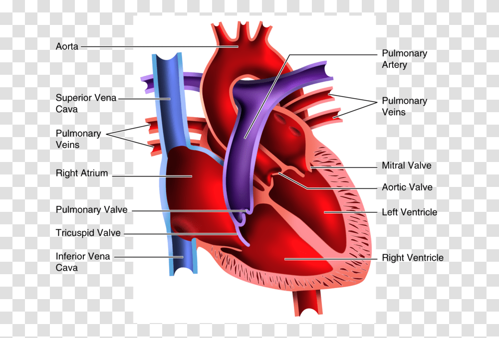
Diagram Of The Human Heart Bs Inferior Vena Cava, Plot ...
Inferior Vena Cava Function, Anatomy & Definition | Body Maps

Congenital absence of inferior vena cava. | Semantic Scholar

Major Blood Vessels Leading to the Heart: Superior Vena Cava, Inferior Vena Cava & Coronary Sinus Video
Inferior vena cava Images, Stock Photos & Vectors | Shutterstock

Anatomy of a Human Heart
0 Response to "40 inferior vena cava diagram"
Post a Comment