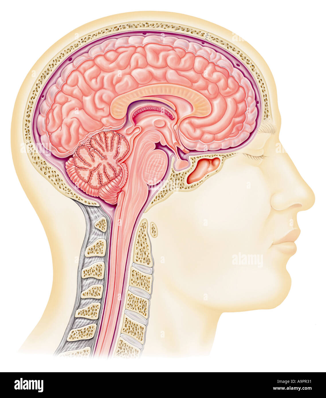
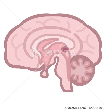
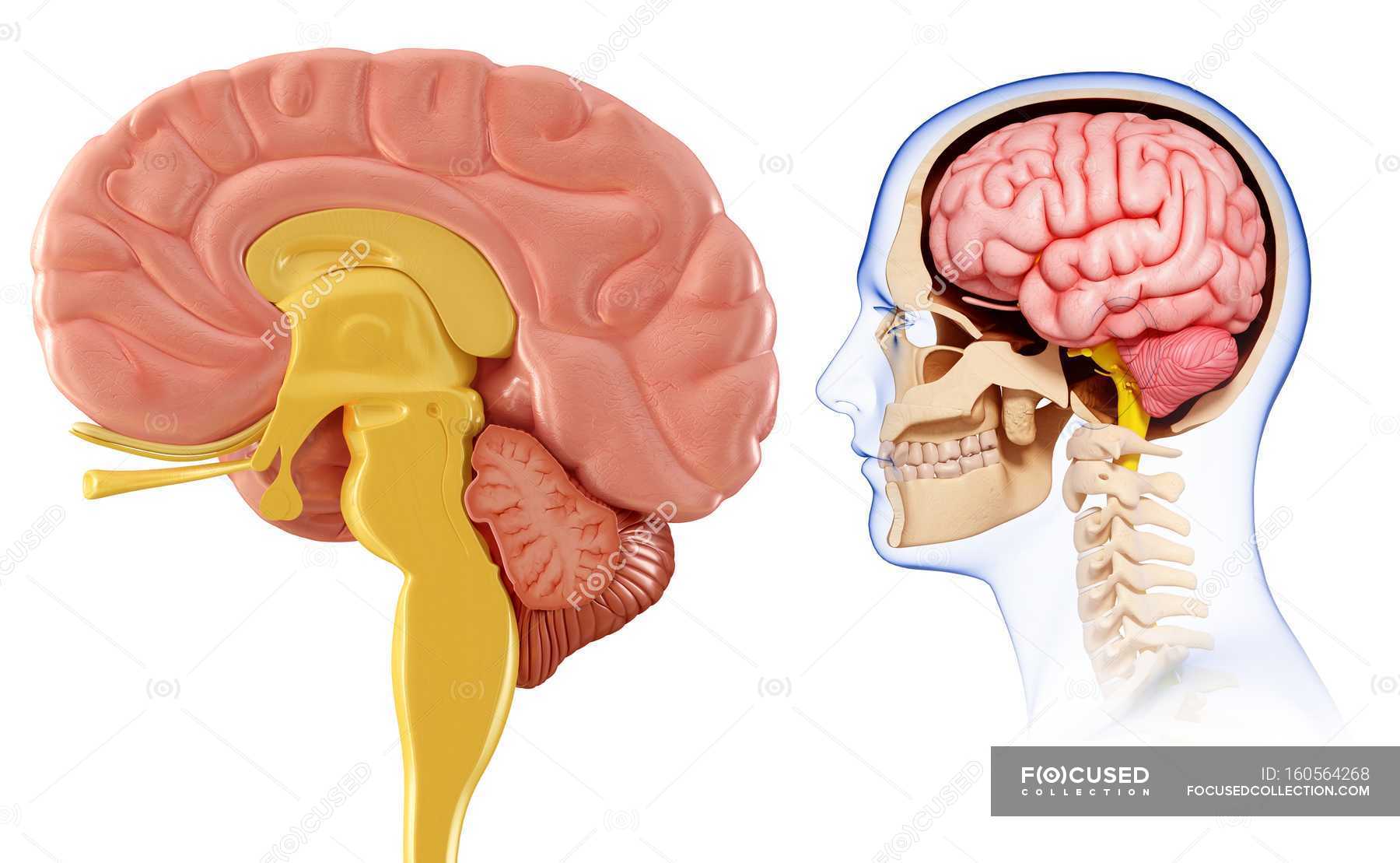
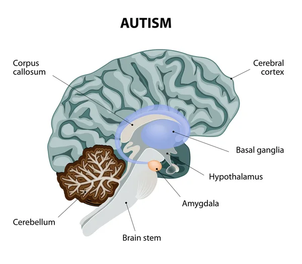
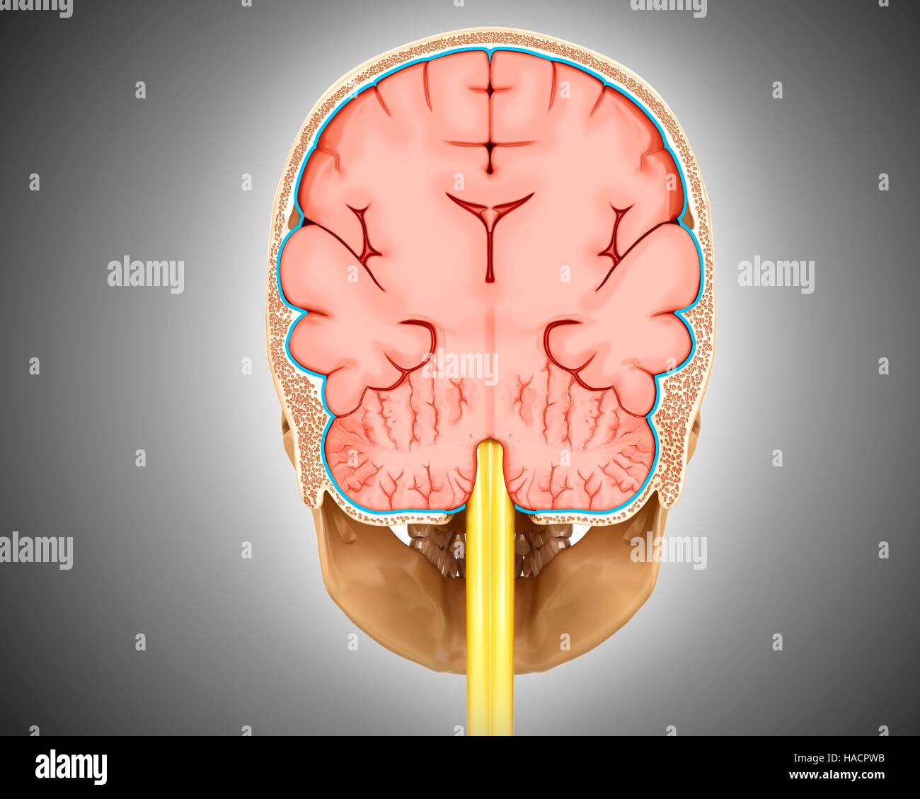
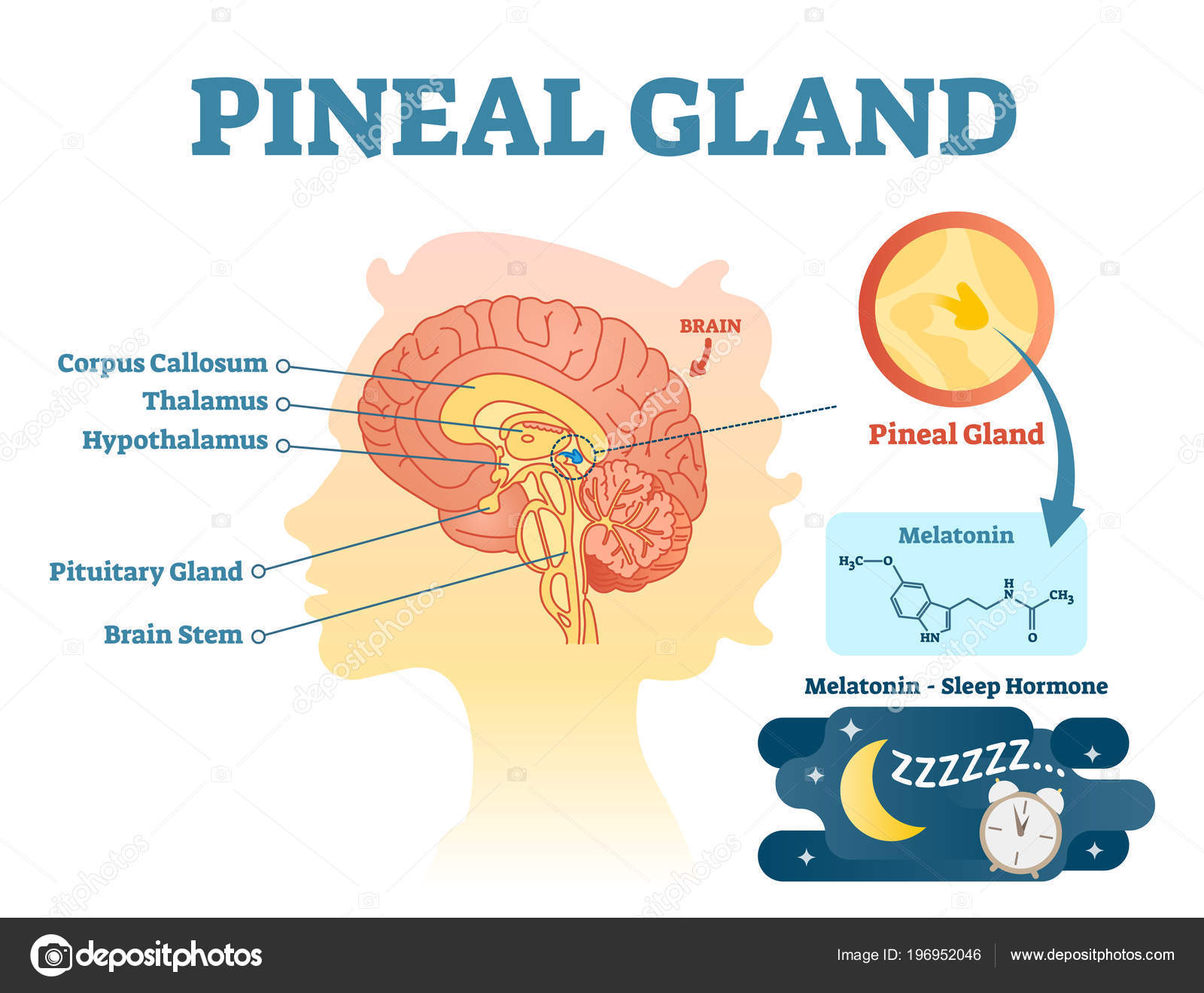
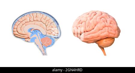
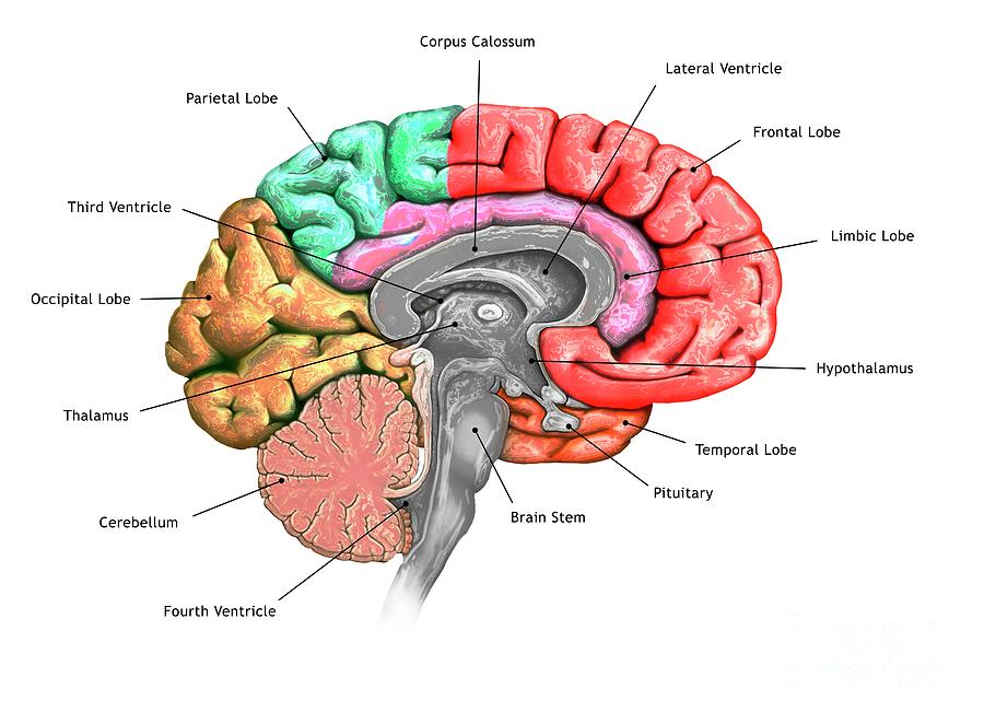
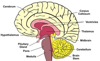
37 brain cross section diagram
If you compare the equations for Q above to the equations for calculating the centroid (discussed in a previous section), you will see that we actually use the first moment of area when calculating the centroidal location with respect to an origin of interest.. The first moment is also used when calculating the value of shear stress at a particular point in the cross section. The principal axis orientation is also indicated on the cross-section diagram. Area: The cross-sectional area of the section. This value is commonly used in determining the axial strength of a column. First Moments of Area: The first moments of area are relevant for certain shear calculations, such as shear flow. Note that the first moments are ...
Cross Section Brain Diagram Brain Diagram Human Brain Nervous System Problems . Cross Section Of The Brain Depicting The Position Occupied By The Anterior Thalamic Nucleus In The Anterior A Nervous System Parts Neuroscience Medical Anatomy .

Brain cross section diagram
BI 335 – Advanced Human Anatomy and Physiology Western Oregon University Figure 4: Mid-sagittal section of brain showing diencephalon (includes corpus callosum, fornix, and anterior commissure) Marieb & Hoehn (Human Anatomy and Physiology, 9th ed.) – Figure 12.10 Exercise 2: Utilize the model of the human brain to locate the following structures / landmarks for the Cross Section Of Human Eye Eps8 Human Brain Anatomy Human Brain Diagram Brain Diagram Brain Parts To Know Brain Diagram Brain Anatomy Human Brain Anatomy I Pinimg Com Originals 66 29 85 66298598bbbcc87 Brain Structures And Functions Worksheet Worksheets Are Obviously The Spine To Scholars Getting To Know And G In 2021 Brain Anatomy Brain […] From the case: Brainstem cross-sectional anatomy (diagrams) Diagram. Entire brainstem. Upper midbrain. From the case: Brainstem cross-sectional anatomy (diagrams) Diagram. Upper midbrain. Lower midbrain. From the case: Brainstem cross-sectional anatomy (diagrams)
Brain cross section diagram. 1,829 cross section of brain stock photos, vectors, and illustrations are available royalty-free. See cross section of brain stock video clips. of 19. brain section human brain cross section human brain cross brain basal ganglia ventricles in the brain spinal reflex basal ganglia cross-section of brain parts of brain cross section of the human ... Cross sectional anatomy: MRI of the brain. An MRI was performed on a healthy subject, with several acquisitions with different weightings: spin-echo T1, T2 and FLAIR, T2 gradient-echo, diffusion, and T1 after gadolinium injection. We obtained 24 axial slices of the normal brain. Data and DICOM images archived on our PACS (Picture Archiving and ... Start studying Brain Cross Section 6. Learn vocabulary, terms, and more with flashcards, games, and other study tools. Cross section through the thalamus: Diagram Orienting yourself within such a cross section is easy. The star of the show (brain) is easily recognizable because it appears highly convoluted, full of ridges (gyri) and indentations (sulci).The paired thalami appear as two circular masses in the midline, forming the walls of the third ventricle.The neurocranium appears as a meshwork (trabecular ...
From the case: Brainstem cross-sectional anatomy (diagrams) Diagram. Entire brainstem. Upper midbrain. From the case: Brainstem cross-sectional anatomy (diagrams) Diagram. Upper midbrain. Lower midbrain. From the case: Brainstem cross-sectional anatomy (diagrams) Cross Section Of Human Eye Eps8 Human Brain Anatomy Human Brain Diagram Brain Diagram Brain Parts To Know Brain Diagram Brain Anatomy Human Brain Anatomy I Pinimg Com Originals 66 29 85 66298598bbbcc87 Brain Structures And Functions Worksheet Worksheets Are Obviously The Spine To Scholars Getting To Know And G In 2021 Brain Anatomy Brain […] BI 335 – Advanced Human Anatomy and Physiology Western Oregon University Figure 4: Mid-sagittal section of brain showing diencephalon (includes corpus callosum, fornix, and anterior commissure) Marieb & Hoehn (Human Anatomy and Physiology, 9th ed.) – Figure 12.10 Exercise 2: Utilize the model of the human brain to locate the following structures / landmarks for the

Human Brain Cross Section Schematic Graphic Illustration Stock Vector Vector And Low Budget Royalty Free Image Pic Esy 043335277 Agefotostock
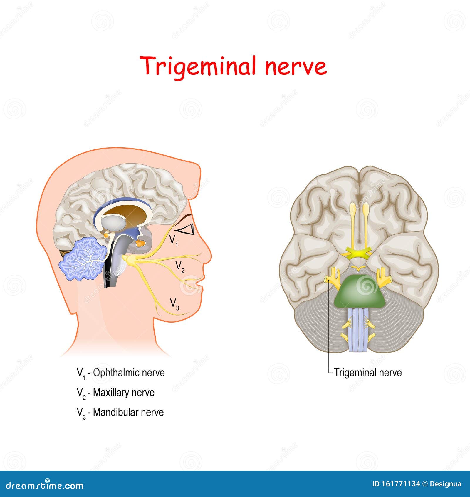
Trigeminal Nerve Head Cross Section And Bottom View Of The Brain Stock Vector Illustration Of Neuralgia Ophthalmic 161771134

Eye Anatomy Blank Worksheet Printable Test Illustrations Brain And Eye Anatomical Cross Section Diagrams School Lesson Activity Educational Workbook Page Learn Human Anatomy Visual Task Poster Id 316139170

Brain Cross Section Diagram Cross Section Of The Human Brain Royalty Free Cliparts Vectors Brain Diagram Human Brain Human Brain Diagram
Draw The Given Diagram Of Cross Sectional View Of Human Brain On Your Answer Sheet And Label Sarthaks Econnect Largest Online Education Community

Amazon Com Functional Human Brain Model Cross Section 1 2 Size Color Coded Numbered With Key Card Includes Mount Eisco Labs Industrial Scientific

The Brain In Cross Section Showing The Basal Ganglia Hypothalamus Amygdala And Hippocampus Royalty Free Cliparts Vectors And Stock Illustration Image 46940834

Cross Section Of The Brain Labeled Anatomy Of Inner Brain Human Anatomy Chart Anatomiya Cheloveka Anatomiya
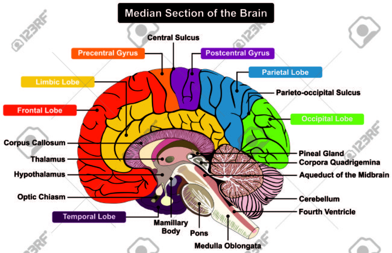
Median Section Of Human Brain Anatomical Structure Diagram Infographic Chart With All Parts Cerebellum Thalamus Hypothalamus Lobes Central Sulcus Medulla Oblongata Pons Pineal Gland Figure Royalty Free Cliparts Vectors And Stock Illustration












0 Response to "37 brain cross section diagram"
Post a Comment