40 muscles of respiration diagram
The Lungs and Breathing. The space between the outer surface of the lungs and inner thoracic wall is known as the pleural space.This is usually filled with pleural fluid, forming a seal which holds the lungs against the thoracic wall by the force of surface tension.This seal ensures that when the thoracic cavity expands or reduces, the lungs undergo expansion or reduction in size accordingly. The muscles of the human body can be categorized into a number of groups which include muscles relating to the head and neck, muscles of the torso or trunk, muscles of the upper limbs, and muscles of the lower limbs. The human body has three different types of muscles. They include: skeletal muscles, smooth muscles, cardiac muscles.
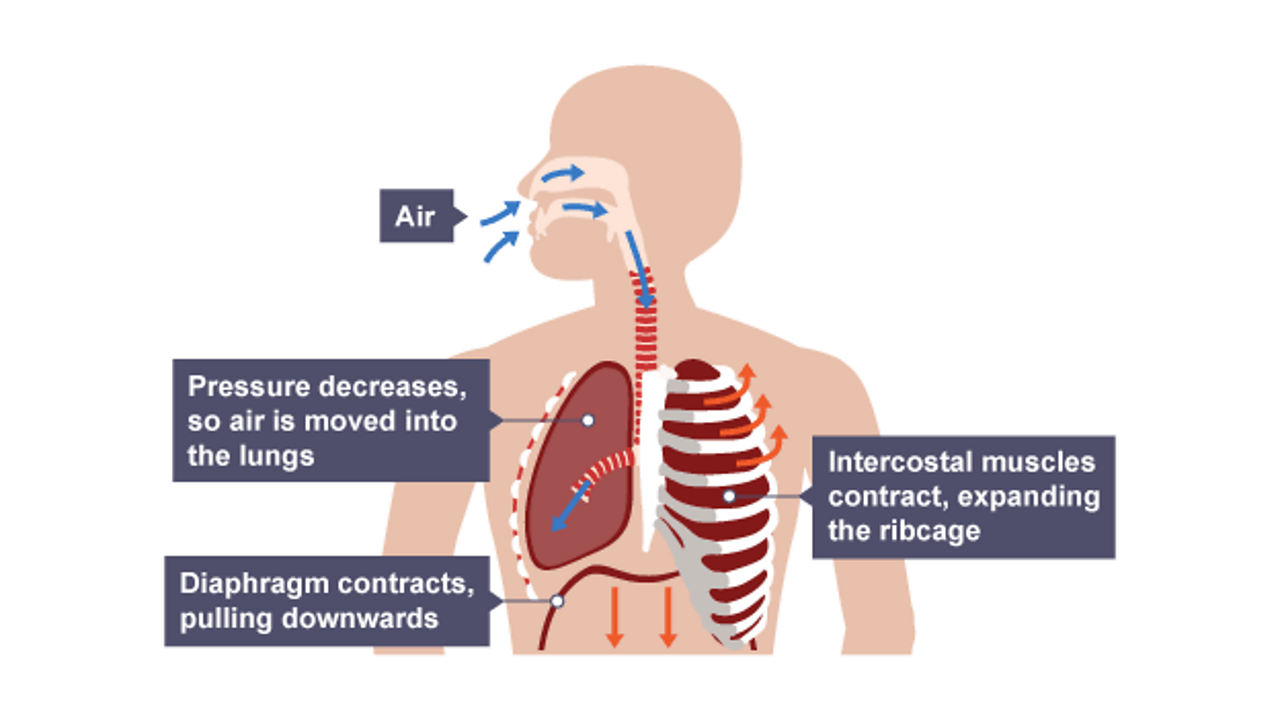
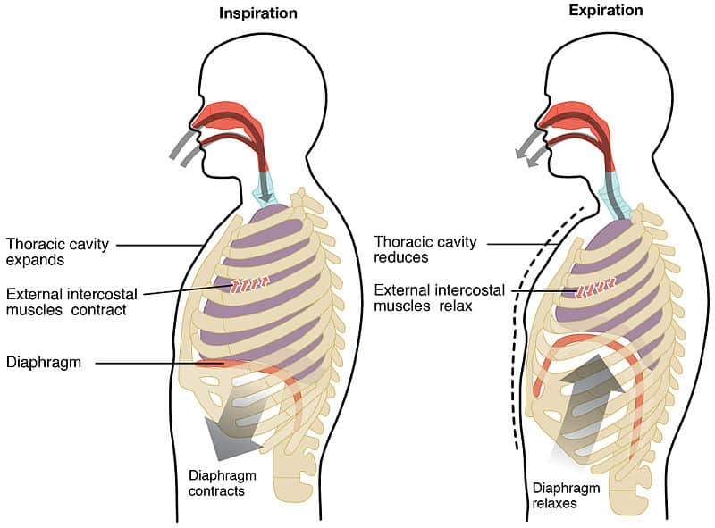
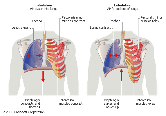
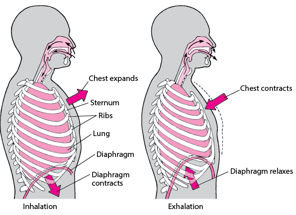
Inspiration - diaphragm contracts and pulls down, intercostal muscles contract and expand the rib cage -> · air enters the lungs · Expiration - diaphragm relaxes ...Breathing cycle: Inspiration - diaphragm contra...Clinical relations: Tachypnea, bradypnea, hyp...Respiratory center: Neuronal groups of the me...Airways: Conducting airways: nose, nasophar...

Muscles of respiration diagram
Muscle Charts of the Human Body For your reference value these charts show the major superficial and deep muscles of the human body. Superficial and deep anterior muscles of upper body Muscles of Respiration. Surrounding the lungs are sets of muscles that are able to cause air to be inhaled or exhaled from the lungs. The principal muscle of respiration in the human body is the diaphragm, a thin sheet of skeletal muscle that forms the floor of the thorax. Steps of cellular respiration. Overview of the steps of cellular respiration. Glycolysis. Six-carbon glucose is converted into two pyruvates (three carbons each). ATP and NADH are made. These reactions take place in the cytosol. Pyruvate oxidation. Pyruvate travels into the mitochondrial matrix and is converted to a two-carbon molecule bound to ...
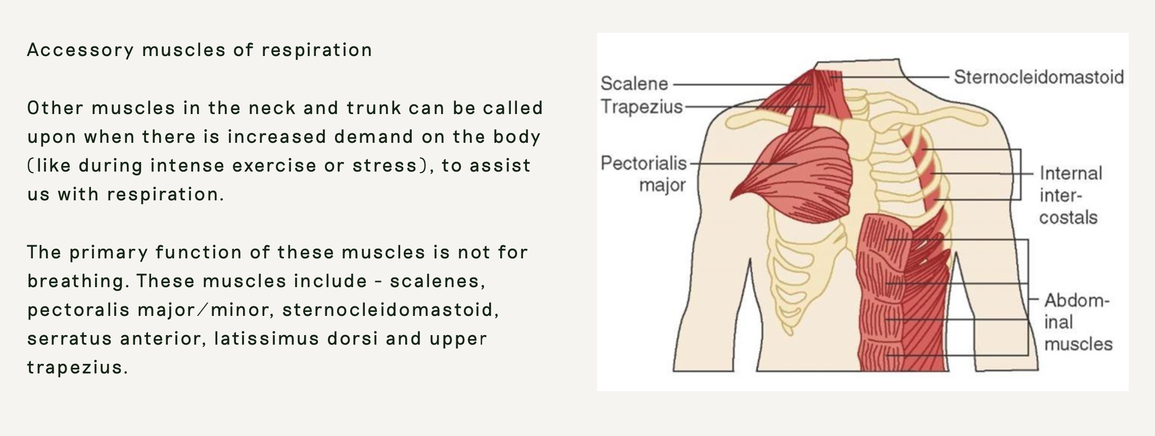
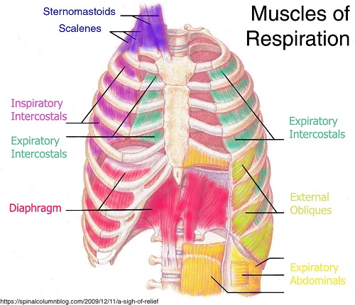
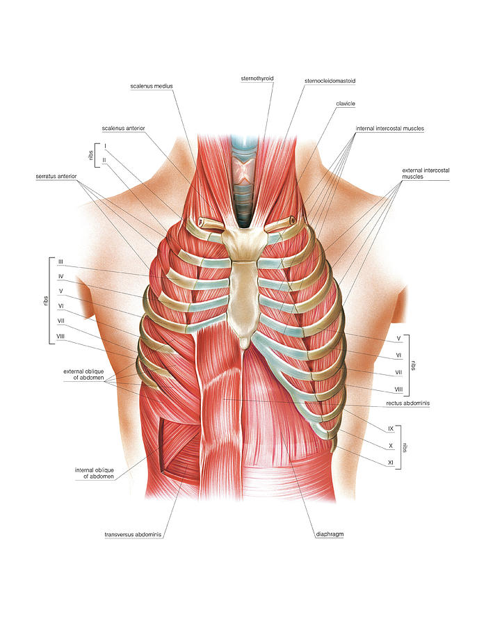
Muscles of respiration diagram. these muscles play a role in lifting up upper part of rib cage, have a small role in respiration scalenus muscle anterior, middle, and posterior muscles of the neck that elevate the first and second ribs The diaphragm and the external intercostals are the muscles primarily responsible for inspiration. Additional muscles are recruited in active respiration. Quiet ...Missing: diagram | Must include: diagram known as anaerobic respiration. Muscles usually receive energy through a process known as cellular respiration[15], but when there is a lack of oxygen in the organism, muscles go through anaerobic respiration. Anaerobic respiration consists of glycolysis with a few extra reactions which allow it to repeat[17]. Glycolysis The breathing muscles include the: Diaphragm, which is a dome-shaped muscle below your lungs. It separates the chest cavity from the abdominal cavity. The diaphragm is the main muscle used for breathing. The muscles between your ribs, called intercostal muscles, play a role in breathing during physical activity.
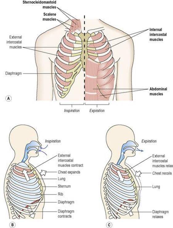
costal cartilages allow movement during inhalation and exhalation. The spaces between the ribs are known as the intercostal spaces and contain intercostal ...1 page The muscles of respiration are also called the 'breathing pump muscles', they form a complex arrangement in the form of semi-rigid bellows around the lungs. All muscles that are attached to the human rib cage have the inherent potential to cause a breathing action. Muscles that helpful in expanding the thoracic cavity are called the inspiratory ... Understanding Cellular Respiration Here are three visual depictions of cellular respiration - an equation, an output description and an illustration. 1) Equation: C 6 H 12 O 6 (1 glucose molecule) + 6 O 2 = 6 CO 2 + 6 H 2 O + 36 ATP (ENERGY) carbohydrate + oxygen = carbon dioxide + water + ATP energy 2) Description of the molecules created in all three stages of cellular respiration: Breathing is an active process - requiring the contraction of skeletal muscles. The primary muscles of respiration include the external intercostal muscles (located between the ribs) and the diaphragm (a sheet of muscle located between the thoracic & abdominal cavities).
The lungs have no skeletal muscles of their own. The work of breathing is done by the diaphragm, the muscles between the ribs (intercostal muscles), ...Missing: diagram | Must include: diagram The scalene muscle group, so important in respiratory dysfunction, is featured in its own article on this site, in the "Perfect Spot" series: Massage Therapy for Neck Pain, Chest Pain, Arm Pain, and Upper Back Pain — Perfect Spot No. 4, an area of common trigger points in the odd scalene muscle group in the neck In general, two muscle groups are used during normal inspiration: the diaphragm and the external intercostal muscles. Additional muscles can be used if a bigger ... Start studying muscles of respiration. Learn vocabulary, terms, and more with flashcards, games, and other study tools.
Muscles Of Respiration Diagram Quizlet
Arm and Hand Muscles Diagram. Muscles of the Chest, Abdomen, Hips, Legs and Feet. ... Back Muscles. Muscles of Respiration. Tendons and Ligaments. Muscle Tone. Characteristics of a Muscle. Growth and Repair of the Muscles. Pathology of the Muscular System. The Skeletal System. Types of Bone. Bone Diagram. Bone Composition. Bones of the Skull ...
The Process Of Breathing Respiration And Gas Exchange Systems Ks3 Biology Bbc Bitesize Bbc Bitesize
Diagram; Steps; Key Points; Respiration is of two types, aerobic respiration, and anaerobic respiration. Aerobic Respiration: It is the process of cellular respiration that takes place in the presence of oxygen gas to produce energy from food. This type of respiration is common in most of the plants and animals, birds, humans, and other mammals.

Respiratory Muscles Quiz
What muscles are used in the expiratory process?
The Respiratory System Ross And Wilson Anatomy And Physiology In Health And Illness 11e
The diaphragm is a dome-shaped sheet of muscle located below the lungs. It separates the chest from the abdomen. The diaphragm operates as the major muscle of respiration and aids breathing .
Anatomy Of Breathing Process And Muscles Of Respiration Kenhub
Respiratory Muscles Diagram 22 November 2018 Edit. Startpage. Respiration Cynical Anatomy Muscles Of Respiration. Solved Questions 3 9identify The Labeled Respiratory Mus Expiration Muscles Abdominal Muscles Internal Intercostal Muscles 2015 Pearson Education Inc Ppt Download
1
Diagram of the Human Respiratory System (Infographic) By Ross Toro 29 August 2013. ... The diaphragm is a dome-shaped muscle below the lungs that controls breathing. The diaphragm flattens out and ...

Pb S Biology Muscles Involved In Breathing External Facebook
Respiratory. The respiratory system, which includes air passages, pulmonary vessels, the lungs, and breathing muscles, aids the body in the exchange of gases between the air and blood, and between ...

Airway Anatomy And Physiology Clinical Essentials Paramedic Care Part 3
ADVERTISEMENTS: The expansion of the lungs during inspiration follows the enlargement of the thoracic cavity in all its diameters by the contraction of the respiratory muscles. These are mentioned below: 1. Diaphragm: A dome-shaped muscle separating the abdominal wall from the thoracic cavity, with vertically running fibres at the periphery and horizontally running fibres at […]

Mechanics And Muscles Of Ventilation Course Hero
The diaphragm is the primary muscle used in respiration, which is the process of breathing. This dome-shaped muscle is located just below the lungs and heart. It contracts continually as you ...
Breathing Good Day Pilates
The respiratory muscles or breathing pump muscles form semi-rigid bellows around the lungs in the chest. It is a complex arrangement of all the muscles which are attached to the rib cage, and help in generating the breathing action. These muscles include inspiratory muscles which cause the thoracic cavity to expand or induce inhalation and ...
Select Chiropractic And Wellness Breathing 101
Muscles get their energy from different sources depending on the situation that the muscle is working in. Muscles use aerobic respiration when we call on them to produce a low to moderate level of force. Aerobic respiration requires oxygen to produce about 36-38 ATP molecules from a molecule of glucose.
Mechanics Of Breathing Inspiration Expiration Teachmephysiology
Muscles of Respiration. During quiet breathing, the predominant muscle of respiration is the diaphragm. As it contracts, pleural pressure drops, which lowers the alveolar pressure, and draws air in down the pressure gradient from mouth to alveoli. Expiration during quiet breathing is predominantly a passive phenomenon, as the respiratory ...

Chapter 10 Respiration During Exercise Ppt Video Online Download
12 photos of the muscle diagram for chest and back. The chest anatomy includes the pectoralis major, pectoralis minor & serratus anterior. The scalenes act as accessory muscles of respiration, and perform flexion at the neck. It is accomplished primarily by the sternocleidomastoid muscles, with assistance.

The Mechanics Of Breathing Phila Massages
The respiratory tract in humans is made up of the following parts: External nostrils - For the intake of air.; Nasal chamber - which is lined with hair and mucus to filter the air from dust and dirt.; Pharynx - It is a passage behind the nasal chamber and serves as the common passageway for both air and food.; Larynx - Known as the soundbox as it houses the vocal chords, which are ...
1
The muscles of respiration are those muscles that contribute to inhalation and exhalation, by aiding in the expansion and contraction of the thoracic cavity.The diaphragm and, to a lesser extent, the intercostal muscles drive respiration during quiet breathing.The elasticity of these muscles is crucial to the health of the respiratory system and to maximize its functional capabilities.

Muscles Of Inspiration Muscles Of Expiration Muscles Of Respiration Respiratory Muscles
Steps of cellular respiration. Overview of the steps of cellular respiration. Glycolysis. Six-carbon glucose is converted into two pyruvates (three carbons each). ATP and NADH are made. These reactions take place in the cytosol. Pyruvate oxidation. Pyruvate travels into the mitochondrial matrix and is converted to a two-carbon molecule bound to ...

Respiratory Anatomy Muscles Of Respiration Diagram Quizlet
Muscles of Respiration. Surrounding the lungs are sets of muscles that are able to cause air to be inhaled or exhaled from the lungs. The principal muscle of respiration in the human body is the diaphragm, a thin sheet of skeletal muscle that forms the floor of the thorax.

C 4 1 Breathing
Muscle Charts of the Human Body For your reference value these charts show the major superficial and deep muscles of the human body. Superficial and deep anterior muscles of upper body

Mechanism Of Breathing And Its Neural Regulation Online Science Notes

Diaphragm Definition Function Location Britannica

Can Tight Muscles Cause Breathing Difficulty Myothrive

The Muscles Of Respiration Netter Illustration Used With Permission Of Download Scientific Diagram
Respiratory Muscles Photograph By Asklepios Medical Atlas

Trunk Muscles Course Hero

Anatomy Of Breathing For Singers Made Easy Healthy Voice With Katarina

The Muscles Of Breathing
Take A Deep Breath James Crader Pilatesintel
Control Of Breathing Lung And Airway Disorders Msd Manual Consumer Version

The Muscles Of Respiration Netter Illustration Used With Permission Of Download Scientific Diagram

Muscles Of Respiration Muscles Of Respiration Muscle Anatomy Anatomy And Physiology Nursing Tips

Respiratory Muscles During Exercise Mechanics Energetics And Fatigue Sciencedirect

Breathing Muscles And Singing Tips For Vocalists

Respiration Muscles

The Muscles Involved In Pulmonary Ventilation Download Scientific Diagram

Mechanics And Muscles Of Ventilation Course Hero

Breathing Monitoring And Pattern Recognition With Wearable Sensors Intechopen

The Physiology Of Respiration
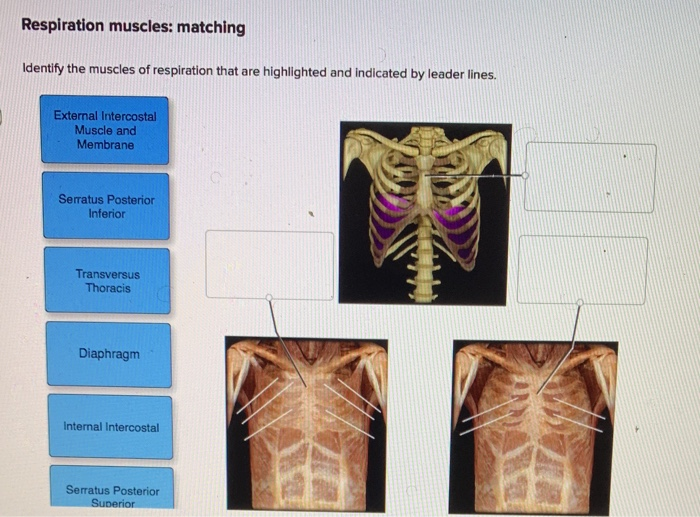
Solved Respiration Muscles Matching Identify The Muscles Of Chegg Com

Ahs 130 Ch 11 Sec 11 5 Muscles Of Respiration Youtube
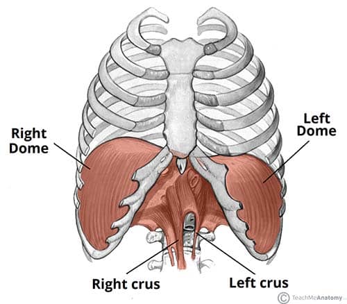
Muscles Of The Thorax Teachmeanatomy
0 Response to "40 muscles of respiration diagram"
Post a Comment