38 Brain And Spinal Cord Diagram
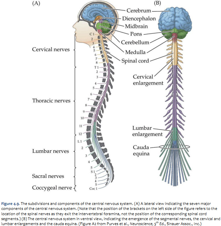
Spinal cord: Anatomy, structure, tracts and function | Kenhub This is the reason why, in adults, the spinal cord occupies only the upper two thirds of the vertebral canal. Spinal cord (diagram) The spinal cord is a continuation of the brainstem. It extends from the foramen magnum at the base of the skull to the L1/L2 vertebra where it terminates as the conus medullaris (medullary cone). Spinal Nerves Function & Diagram | What Are The 31 Pairs ... Spinal Nerves. There are many different organ systems in the human body. One of tehse is the nervous system which is comprised of the brain and spinal cord.
Human Spinal Cord Diagram - 31 spinal cord parts and ... Human Spinal Cord Diagram. Here are a number of highest rated Human Spinal Cord Diagram pictures on internet. We identified it from trustworthy source. Its submitted by dealing out in the best field. We admit this kind of Human Spinal Cord Diagram graphic could possibly be the most trending subject when we allowance it in google plus or facebook.
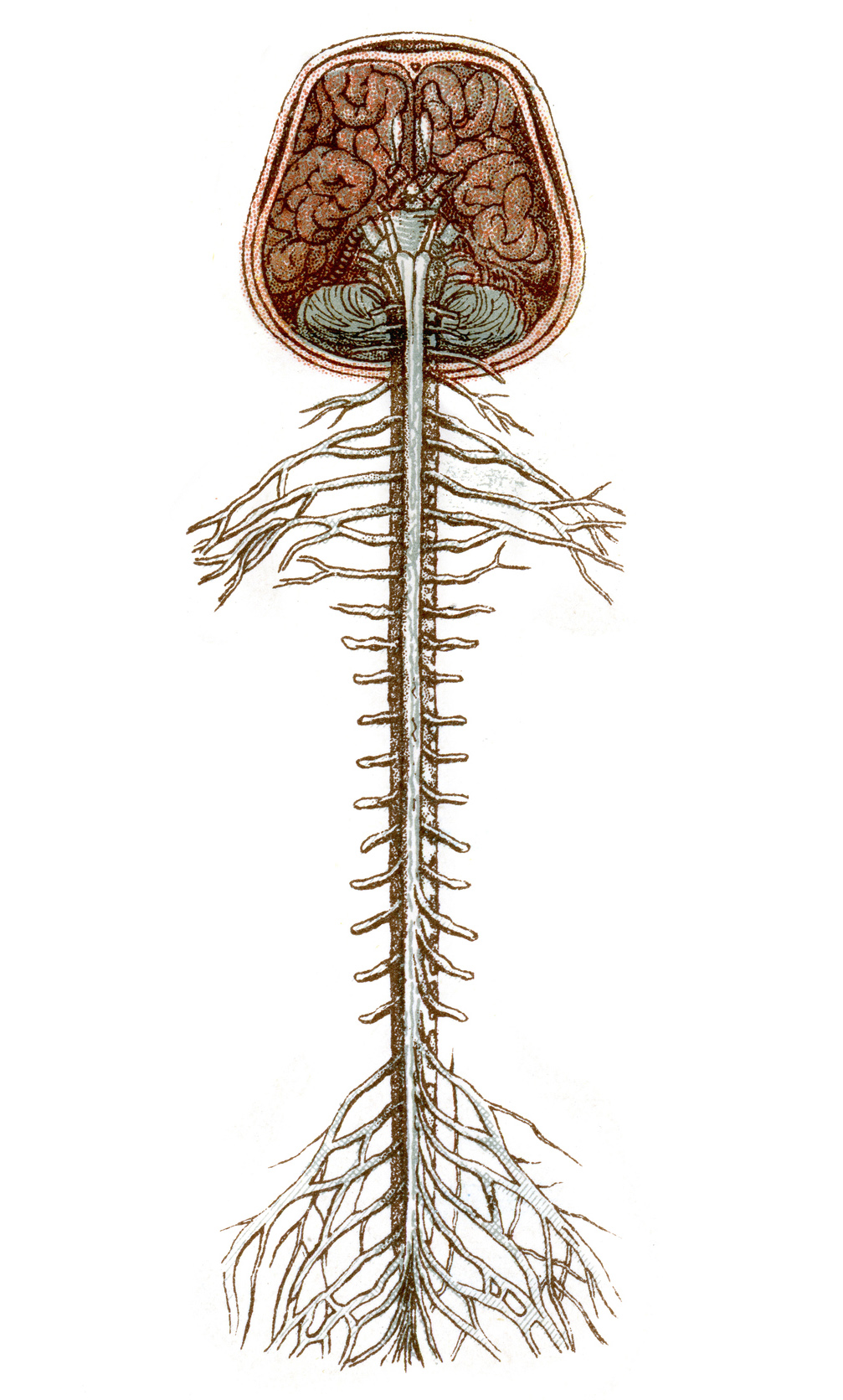
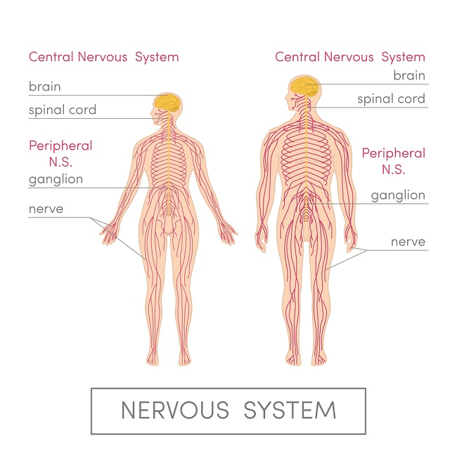
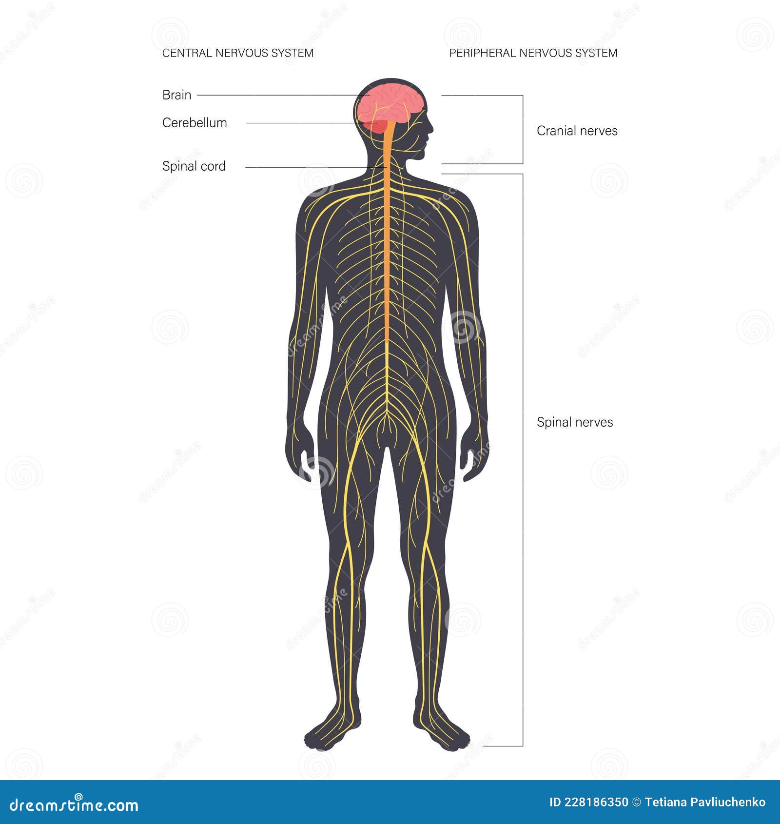
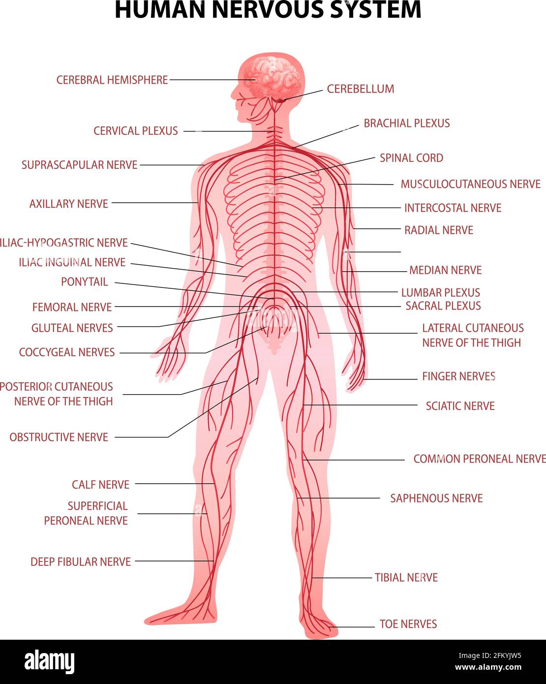
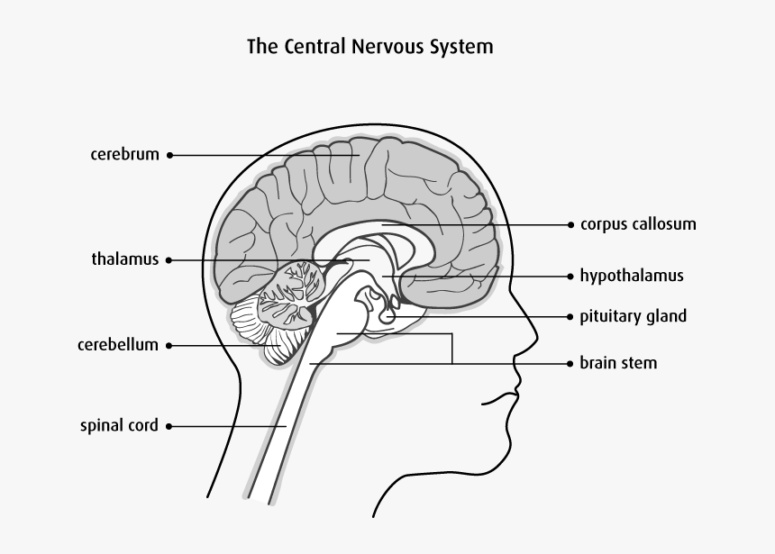
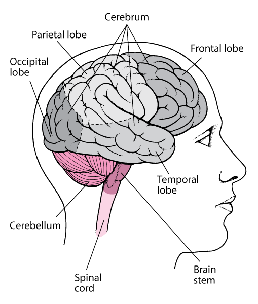
Brain and spinal cord diagram
Brain And Spine Diagram - Studying Diagrams The spinal cord like the brain has two major layers of protection. Finally a diagram summarizes the insertion and origin of the transversal spinalis muscles semispinalis multifidus and rotator muscles. It is situated inside the vertebral canal of the vertebral column. The cerebral cortex is divided lengthways into two cerebral. diagramsketch.com › brain-and-spinal-cord-diagramBrain And Spinal Cord Diagram - Diagram Sketch Dec 17, 2021 · Prop Haphead Interface Brain Diagram Spinal Cord Anatomy Brain Anatomy. The Coverings Of The Brain And Spinal Cord A The Brain And Spinal Cord Are Covered With Thre Human Anatomy And Physiology Nervous System Anatomy Spinal Cord. Pin On Cc Cycle 3. Pin On Teoc. Diagram Of The Central Nervous System Central Nervous System Nervous Nervous System. Anatomy, Head and Neck, Dura Mater - StatPearls - NCBI ... The dura mater often gets referred to as merely the dura. It is one of the layers of connective tissue that make up the meninges of the brain (pia, arachnoid, and dura, from inside to outside). It is the outermost layer of the three meninges that surround and protect the brain and spinal cord. The dura mater is made up of fibroblasts and large amounts of extracellular collagen.[1]
Brain and spinal cord diagram. Interneurons Function, Diagram & Location | Association ... The brain recognizes the situation and sends instructions back down the spinal cord, through interneurons, and on to motor neurons in muscles directing the person to swat the insect off their leg. Learn the spinal cord with diagrams and quizzes | Kenhub The spinal cord, along with the brain, makes up the central nervous system (CNS). It is a long tubular structure comprised of nervous tissue, extending from the cervical to the lumbar region of the vertebral column. Just like other parts of the CNS, the spinal cord is comprised of white and gray matter. Spinal cord gray matter is the central ... The Myelin Sheath: Anatomy, Function, and Treatment Guanabenz has also been shown to reduce the number of inflammatory immune cells that collect in the brain and spinal cord. Ibudilast: A phase 2 trial involving 255 people with primary or secondary progressive MS found that ibudilast, an anti-inflammatory drug made in Japan, slowed the rate of brain atrophy (shrinking) compared to placebo. Diagram Of The Brain And Its Functions - Studying Diagrams Diagram Of The Brain And Its Functions nisa on November 10, 2021 The human brain is the central organ of the human nervous system and with the spinal cord makes up the central nervous system. The forebrain is responsible for controlling emotions hunger sleep body temperature reproductive body parts and functions.
Nerves Of The Brain Diagram - Studying Diagrams Spinal Nerves There are many different organ systems in the human body. Multiple sclerosis MS is a potentially disabling disease of the brain and spinal cord central nervous system. Diagram 145 - The nervous system of a horse. Smaller nerves are grouped into larger rope-like groups that travel up and down the body. Neuroanatomy, Central Nervous System (CNS) - StatPearls ... The spinal cord is responsible for transmitting signals between the periphery and the rest of the CNS through both ascending and descending pathways. Due to unequal growth during development, the spinal cord is shorter than the spinal column, with the cell bodies of the spinal nerves ending around the level of L1/L2 at the conus medullaris. Physiological Processes of the Nervous System | Overview ... The spinal cord runs from the bottom of the brain stem to the lower back and is encased in bone called the spinal column. Nerves all over the body detect and send electrochemical signals, also ... Difference between Brain and Spinal Cord: Definition and ... Check Important Notes for Spinal Cord Diagram: Detailed Illustration The brain mainly is composed of two types of tissues. Grey Matter, which consists of cells that make up the bulk of the brain. White Matter, which is primarily composed of axons, connects various areas of grey area together. Structure and Functions of the Brain
Central Nervous System Diagram Drawing - Central Nervous ... After reading the kidshealth articles related to the brain and nervous system, draw a map of the brain. The nervous system has two main parts: The central nervous system (cns) is the part of the nervous system that includes the brain and spinal cord. We were unable to load the diagram. Music And The Brain Diagram - Studying Diagrams Together the brain and spinal cord that extends from it make up the central nervous system or CNS. But engineers at Google. 13442 human brain diagram stock photos vectors and illustrations are available royalty-free. researchgate.net › figure › Diagram-of-the-brain-andDiagram of the brain and spinal cord. Diagram of the brain ... Diagram of the brain and spinal cord. Diagram of the brain and spinal cord illustrating how the eye’s inner and outer blood-retinal barriers (BRBs) fit into the overall scheme of blood-neural ... Brain Meninges Diagram - Diagram Sketch There Are 3 Layers Of Protection And Support Between The Brain And The Skull The Dura Mater The Arachnoid Lay Nervous System Anatomy Dura Mater Brain Anatomy. The Coverings Of The Brain And Spinal Cord A The Brain And Spinal Cord Are Covered With Thre Human Anatomy And Physiology Nervous System Anatomy Spinal Cord. Leptomeninges.
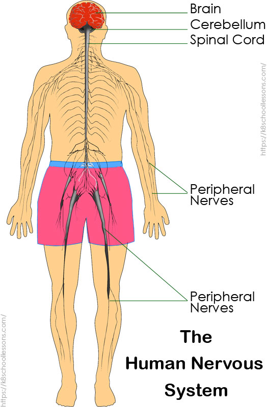
Diy Diagram Of Nervous System - Glaucoma Template The nerves from the brain and the spinal cord connect the skeletal muscles and control their activity according to the directions and demands of the body. Browse 1440 nervous system diagram stock photos and images available or search for central nervous system diagram to find more great stock photos and pictures.
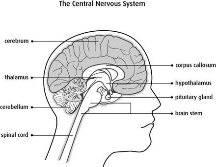
Parts of the Brain Activity for Kids, Brain Diagram, and ... Parts of the Brain Activity for Kids, Brain Diagram, and Worksheets for Kids. August 3, 2021. ... BRAIN STEM - The brain stem connects the rest of the brain to the spinal cord, which runs down your neck and back. The brain stem is in charge of all the functions your body needs to stay alive, like breathing air, digesting food, and circulating ...
Diagram Of Meninges Of Brain - Diagram Sketch Diagram Of Meninges Of Brain. Schematic Of The Meninges And Their Relationships To The Brain And Skull Subdural Hematoma Intracranial Pressure Cerebrospinal Fluid. Brain Anatomy The Function And Layers Of The Meninges Brain Anatomy Nervous System Anatomy Basic Anatomy And Physiology. The Coverings Of The Brain And Spinal Cord A The Brain And ...
Meninges: Dura, arachnoid, pia, meningeal spaces - Kenhub Meninges of the brain. The meninges are the three membranes that envelop the brain and spinal cord and separate them from the walls of their bony cases (skull and vertebral column).Based on their location, meninges are referred to as the cranial meninges which envelop the brain, and spinal meninges which envelop the spinal cord. However, the cranial and spinal meninges are continuous with each ...
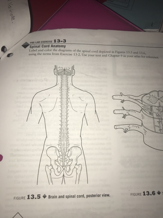
Anatomy of the spinal cord - eAnatomy This atlas of human anatomy describes the spinal cord through 18 anatomical diagrams with 270 anatomical structures labeled. It was designed particularly for physiotherapists, osteopaths, rheumatologists, neurosurgeons, orthopedic surgeons and general practitioners, especially for the study and understanding of medullary diseases.
byjus.com › biology › spinal-cord-diagramSpinal Cord Diagram with Detailed Illustrations and Clear Labels Spinal Cord Diagram. The spinal cord is one of the most important structures in the human body. In fact, it is the most important structure for any vertebrates. Anatomically, the spinal cord is made up is made up of nervous tissue and is integrated into the spinal column of the backbone.
Awesome Central Nervous System Diagram - Glaucoma Template The central nervous system is composed of the brain and the spinal cord. Here is a diagram that you can refer to while you read about the human nervous system function and parts. The CNS receives input from a variety of different sources and implements an appropriate response to the stimuli in a cohesive manner.
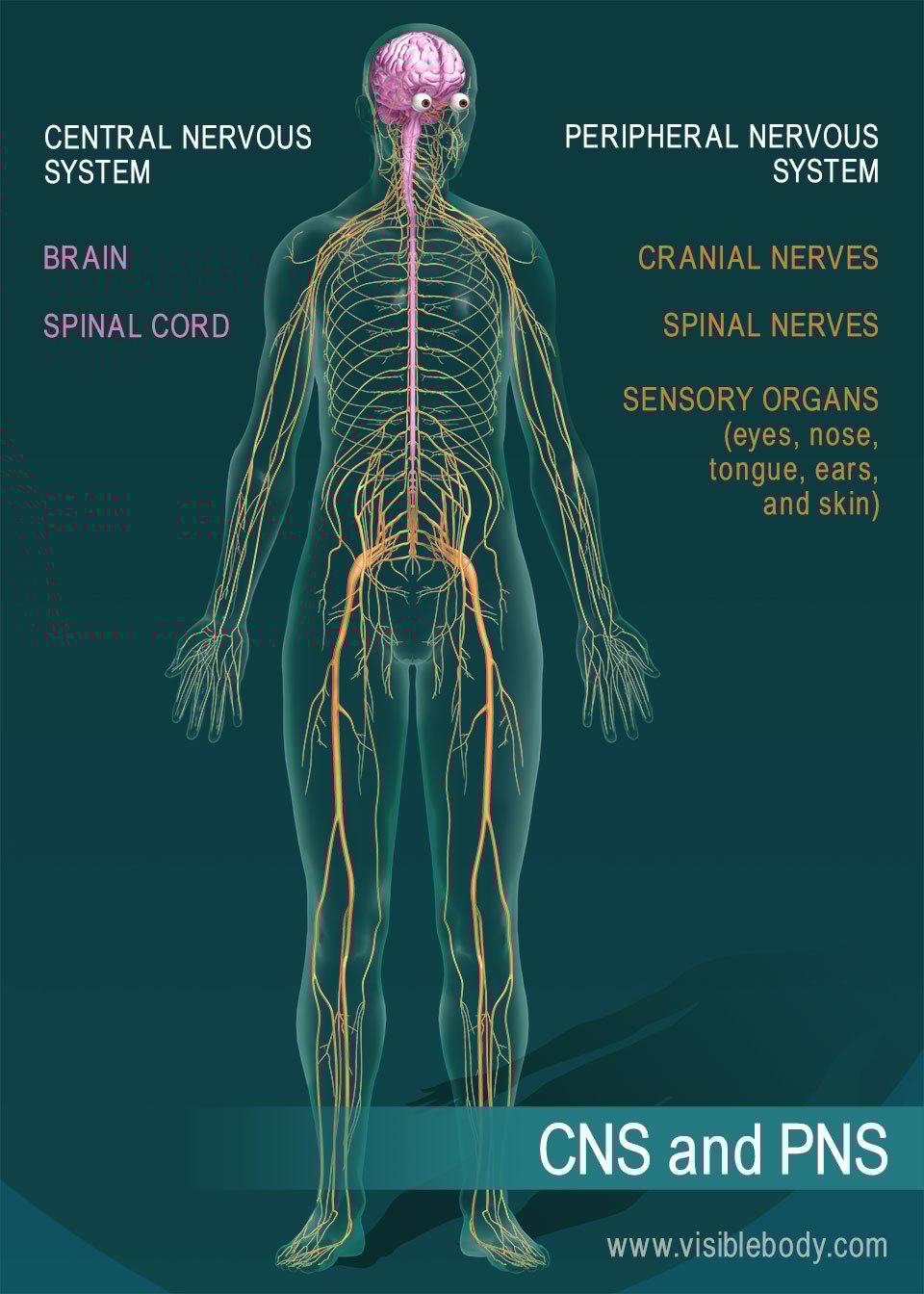
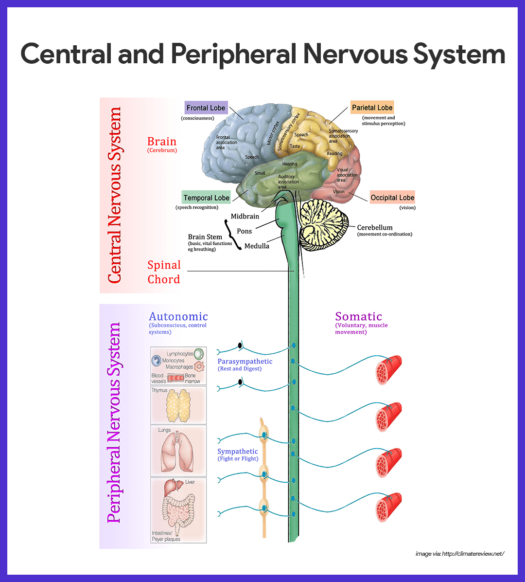
Central nervous system: Anatomy, structure, function | Kenhub The brain consists of the cerebrum, subcortical structures, brainstem and cerebellum. The spinal cord continues inferiorly from the brainstem and extends through the vertebral canal. While analyzing the information and preparing adequate body responses, the parts of the brain and spinal cord communicate with each other via many neural pathways.
35.3: The Central Nervous System - Biology LibreTexts The central nervous system (CNS) is made up of the brain, a part of which is shown in Figure 35.3. 1 and spinal cord and is covered with three layers of protective coverings called meninges (from the Greek word for membrane). The outermost layer is the dura mater (Latin for "hard mother").
Nervous system: Structure, function and diagram | Kenhub Spinal nerves (diagram) Spinal nervesemerge from the segments of the spinal cord. They are numbered according to their specific segment of origin. Hence, the 31 pairs of spinal nerves are divided into 8 cervical pairs, 12 thoracic pairs, 5 lumbar pairs, 5 sacral pairs, and 1 coccygeal spinal nerve. All spinal nerves are mixed, containing both ...
studyingdiagrams.com › brain-and-spinal-cord-diagramBrain And Spinal Cord Diagram - Studying Diagrams Jan 28, 2022 · The brain is one of the most complex and magnificent organs in the human body. The white matter of the spinal cord consists of myelin which performs the function of electrical insulation. The cerebrum functions in. Spinal Cord Diagram. The brain stem begins inferior to the thalamus and runs approximately 7 cm before merging into the spinal cord.
Free Nervous System Worksheets and Printables The nervous system is a complex network of nerves and cells that transmit messages to two main parts of our body: the brain and spinal cord. Human Nervous System The human nervous system is the part of human anatomy that sends signals from sensory receptors to the spinal cord and brain and then transmits the impulses back to the other parts of ...
Brain: Function and Anatomy, Conditions, and Health Tips The brain stem is located in front of the cerebellum and connects to the spinal cord. It's responsible for passing messages to various parts of the body and the cerebral cortex. It consists of ...
Anatomy, Head and Neck, Dura Mater - StatPearls - NCBI ... The dura mater often gets referred to as merely the dura. It is one of the layers of connective tissue that make up the meninges of the brain (pia, arachnoid, and dura, from inside to outside). It is the outermost layer of the three meninges that surround and protect the brain and spinal cord. The dura mater is made up of fibroblasts and large amounts of extracellular collagen.[1]
diagramsketch.com › brain-and-spinal-cord-diagramBrain And Spinal Cord Diagram - Diagram Sketch Dec 17, 2021 · Prop Haphead Interface Brain Diagram Spinal Cord Anatomy Brain Anatomy. The Coverings Of The Brain And Spinal Cord A The Brain And Spinal Cord Are Covered With Thre Human Anatomy And Physiology Nervous System Anatomy Spinal Cord. Pin On Cc Cycle 3. Pin On Teoc. Diagram Of The Central Nervous System Central Nervous System Nervous Nervous System.
Brain And Spine Diagram - Studying Diagrams The spinal cord like the brain has two major layers of protection. Finally a diagram summarizes the insertion and origin of the transversal spinalis muscles semispinalis multifidus and rotator muscles. It is situated inside the vertebral canal of the vertebral column. The cerebral cortex is divided lengthways into two cerebral.
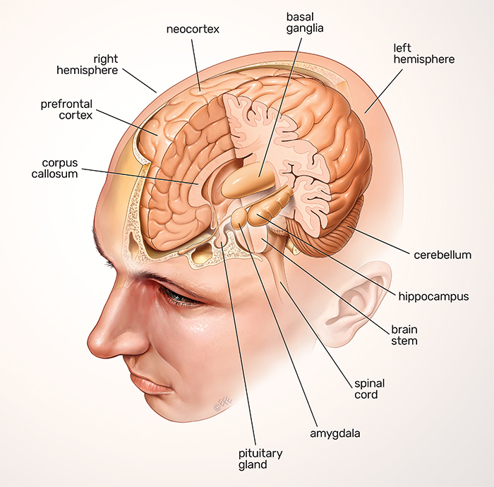
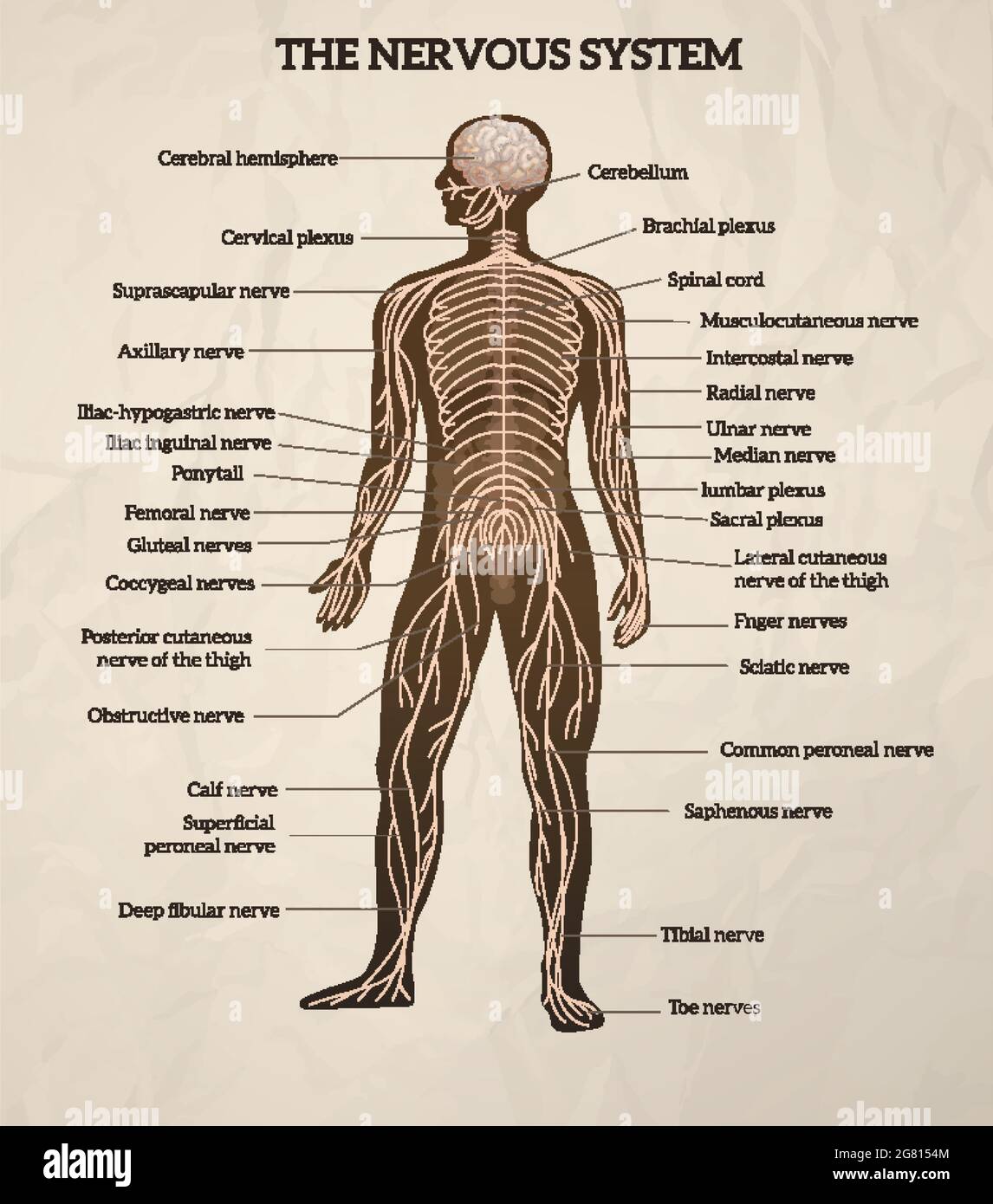

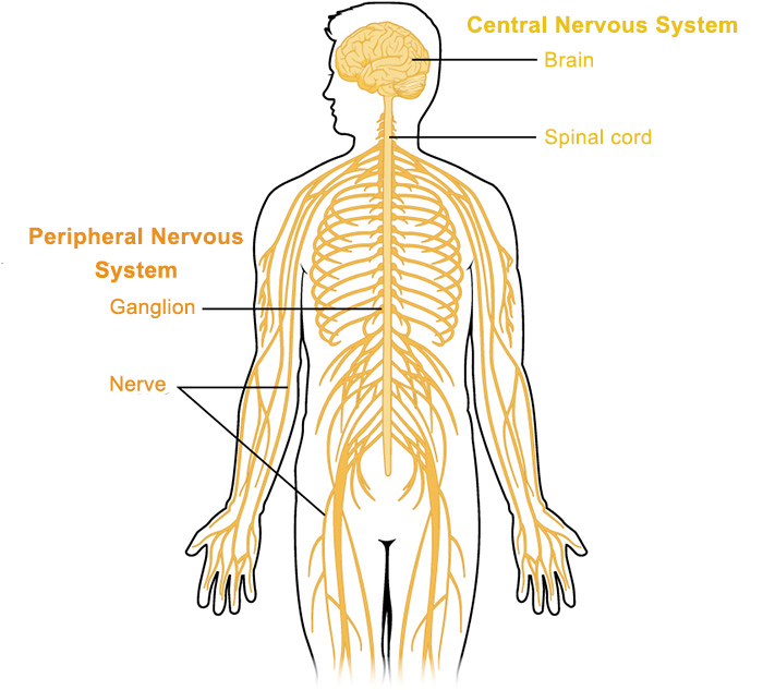


(246).jpg)








:background_color(FFFFFF):format(jpeg)/images/library/12660/anatomy-vertebral-column_-b_english.jpg)


![spinal_cord [Operative Neurosurgery]](https://operativeneurosurgery.com/lib/exe/fetch.php?tok=922dc3&media=http%3A%2F%2Fwww.daviddarling.info%2Fimages%2Fspinal_cord.jpg)

0 Response to "38 Brain And Spinal Cord Diagram"
Post a Comment