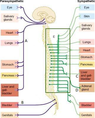
39 drag the labels onto the diagram to identify the components of the autonomic nervous system.
Solved Drag the labels onto the diagram to identify the - Chegg Transcribed image text: Prag the labels onto the diagram to identify the components of the autonomic nervous system! Reset Help Cardiac muscle Smooth muscle ...1 answer · Top answer: Visceral motor nuclei in hypothalamus. Smooth muscle. G... drag the labels into the diagram to identify the structures ... Drag the labels onto the diagram to identify the components of the autonomic nervous system. Prag the labels onto the diagram to identify the components of ...1 answer · 0 votes: [ Lateral forn Lateral Horn Isplanchic Nerve Post ganglionic Twhite Ramey Communion fbry Collateral ganglion
Anatomy, Autonomic Nervous System - StatPearls - NCBI ... The autonomic nervous system is a component of the peripheral nervous system that regulates involuntary physiologic processes including heart rate, blood pressure, respiration, digestion, and sexual arousal. It contains three anatomically distinct divisions: sympathetic, parasympathetic, and enteric.
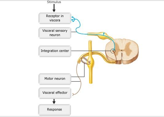
Drag the labels onto the diagram to identify the components of the autonomic nervous system.
EOF What Is Negative Feedback Loop of ... - New Health Advisor Homeostasis is disrupted when one of the key parts of the system isn't working right or something is off. The body then goes through a series of changes to try and bring things back into balance. For example, when you get cold, you shiver to bring up your temperature. All of these responses to change the body start in the brain and gets sent ... 44 drag the labels onto the diagram of muscle spindle ... Solved Drag the labels onto the diagram of muscle spindle Transcribed image text: Drag the labels onto the diagram of muscle spindle function. Reset Muscle shortens Strelch receptors in the spinde are activated throughout the contraction Muscle length Gamma mator neurons activate intrafusal fibers Intrafusal fibers do not slacken so firing rate ...
Drag the labels onto the diagram to identify the components of the autonomic nervous system.. Peripheral nervous system: Anatomy, divisions, functions ... A nervous system division composed of all the neural tissue found outside the cranial vault and vertebral canal. Anatomical components. Peripheral nerves (spinal nerves, cranial nerves, autonomic nerves) Ganglia. Functional components. Autonomic nervous system (ANS) - involuntary part in control of cardiac, smooth and glandular cells. The Cervical Plexus - Spinal nerves - TeachMeAnatomy The cervical plexus is a network of nerve fibres that supplies innervation to some of the structures in the neck and trunk.. It is located in the posterior triangle of the neck, halfway up the sternocleidomastoid muscle, and within the prevertebral layer of cervical fascia. The plexus is formed by the anterior rami (divisions) of cervical spinal nerves C1-C4. The Renin-Angiotensin-Aldosterone-System - TeachMePhysiology The Renin-Angiotensin-Aldosterone System (RAAS) is a hormone system within the body that is essential for regulation of blood pressure and fluid balance. It is comprised of the three hormones renin, angiotensin II and aldosterone and regulated primarily by renal blood flow. This article shall discuss the system, how it is regulated and clinically relevant conditions to its dysfunction. Anatomy & Physiology: BIO 161 / 162 - Community College of ... Chapter 13: Anatomy of the Nervous System ; Chapter 14: The Somatic Nervous System ; Chapter 15: The Autonomic Nervous System ; Chapter 16: The Neurological Exam ; Chapter 17: The Endocrine System ; AP 1 Lab. AP 1 Lab Atlas ; AP 1 Full Lab Manual ; Lab 1: Introduction, Body Organization, and Organ Systems ; Lab 2: Tissues ; Lab 3: Integumentary ...
Autonomic nervous system: Anatomy, divisions, function ... The autonomic nervous system (ANS) is a functional division of the nervous system, with its structural parts in both the central nervous system (CNS) and the peripheral nervous system (PNS). It controls the glands and smooth muscle of all the internal organs (viscera) unconsciously. This is why it's also called the visceral nervous system. Answered: Part B Drag the labels onto the diagram… | bartleby Solution for Part B Drag the labels onto the diagram to identify structural features associated with the extrinsic muscles that move the foot and toes.1 answer · Top answer: Step 1 Introduction: a. Fibularis longus extends from proximal side of fibula and reaches first metatarsal and medial cuneiform bone. This muscle is responsible ... 44 drag the labels onto the diagram of muscle spindle ... Solved Drag the labels onto the diagram of muscle spindle Transcribed image text: Drag the labels onto the diagram of muscle spindle function. Reset Muscle shortens Strelch receptors in the spinde are activated throughout the contraction Muscle length Gamma mator neurons activate intrafusal fibers Intrafusal fibers do not slacken so firing rate ... What Is Negative Feedback Loop of ... - New Health Advisor Homeostasis is disrupted when one of the key parts of the system isn't working right or something is off. The body then goes through a series of changes to try and bring things back into balance. For example, when you get cold, you shiver to bring up your temperature. All of these responses to change the body start in the brain and gets sent ...
EOF








0 Response to "39 drag the labels onto the diagram to identify the components of the autonomic nervous system."
Post a Comment