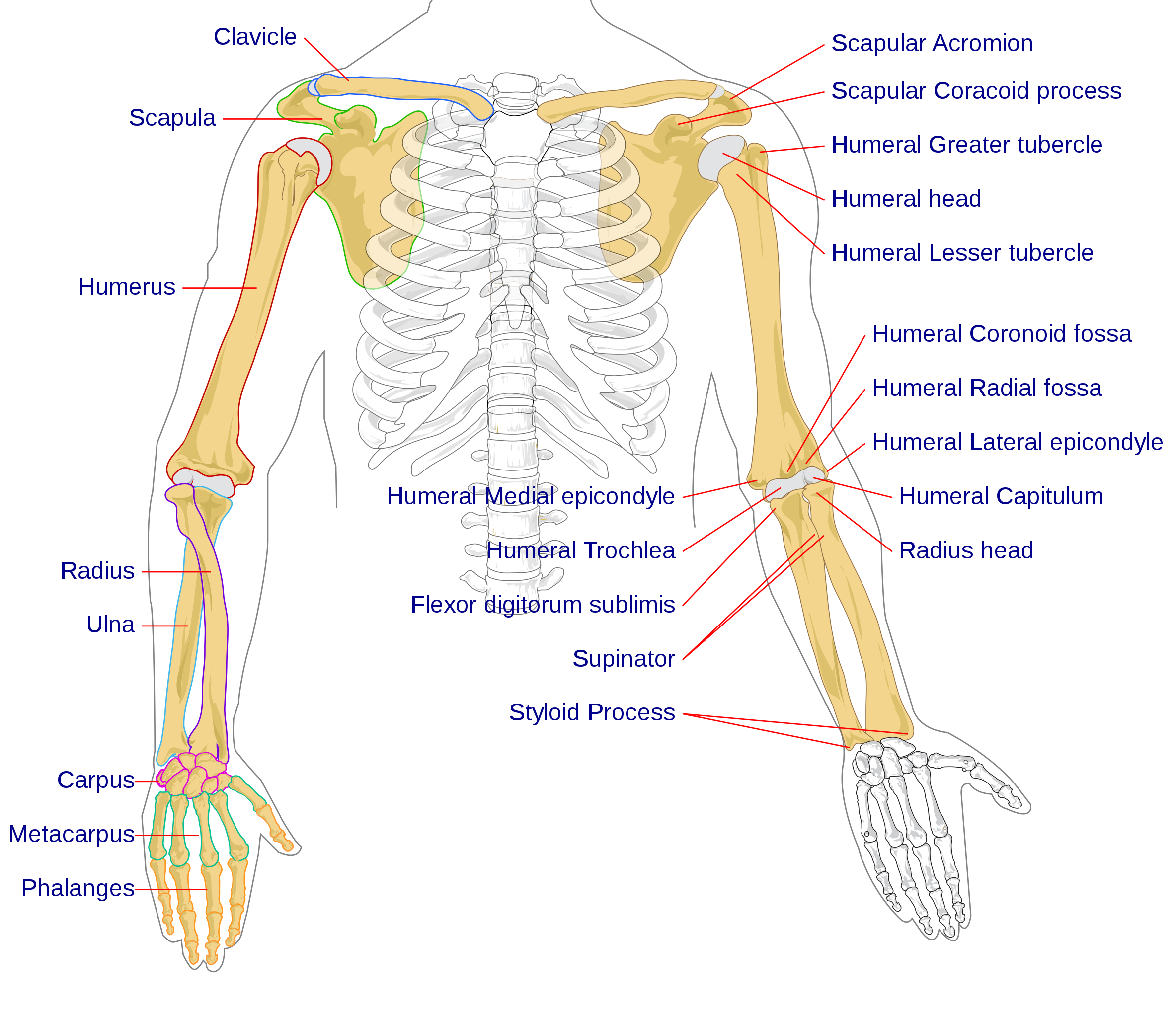
37 skeletal system diagram pdf
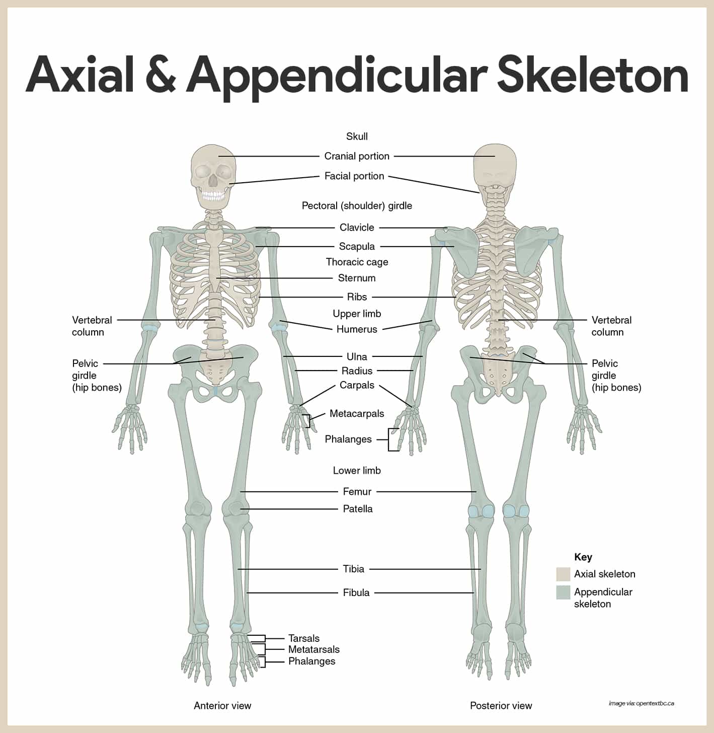
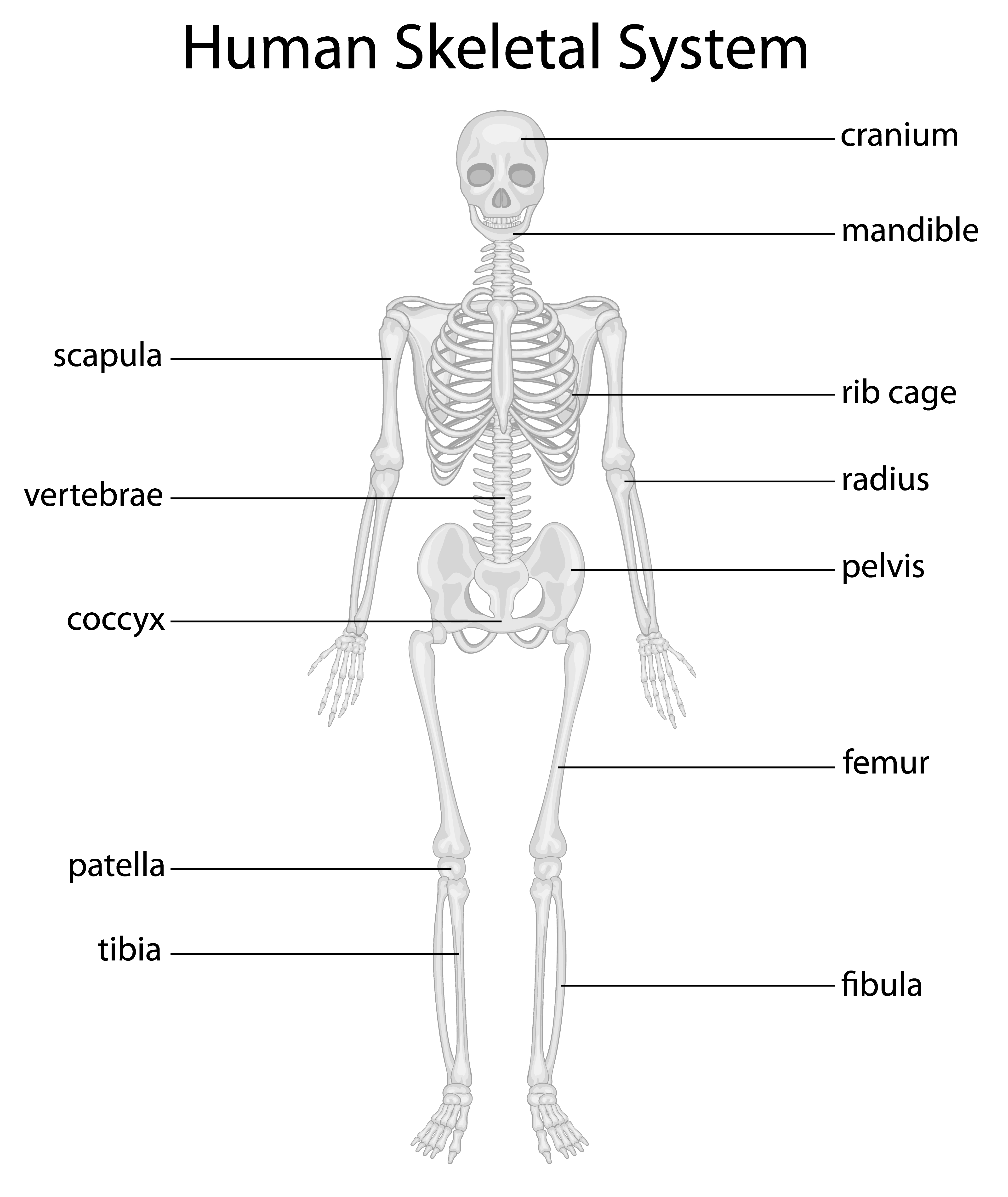
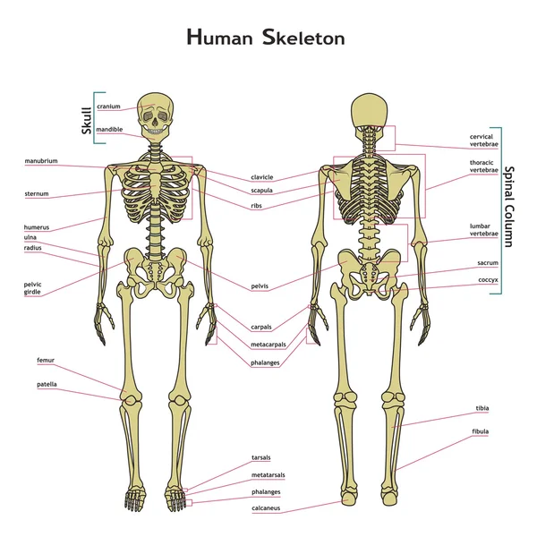
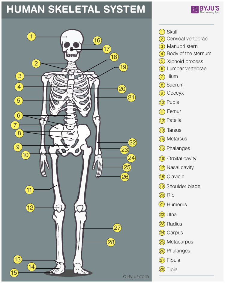
types of bones—each with different functions—work together to give the skeletal system strength and effectiveness. Long bones are cylindrical in shape and consist of a shaft with two ends. Found in limbs, long bones typically support body weight and act as levers. The femur, or thighbone, is an example of a long bone.Short bones are cube-like in that they are nearly equal in length and ... Skeletal system diagrams are illustrations of the human skeleton, used mostly for educational purposes or in presentations. Skeletal diagrams are tools used by students to learn and study all 206 bones (this number can vary from person to person) of the human body. There are four main categories of bones: long, short, irregular, and flat.
Bones-r-us. The skeleton is divided into two parts: the axial (from the Latin axis, meaning axle or wheel) and appendicular (from the Latin appendare, ...15 pages

Skeletal system diagram pdf
To be able to grow, strong bones needs lots of calcium and weight-bearing physical activity. Side of skull. (Parietal bone)2 pages The skeletal system consists of 206 bones that make up the internal framework of the body, called the skeleton. The skeleton supports the body, protects internal organs, serves as a point of attachment for skeletal muscles for body movement, produces blood cells, and stores minerals. Organs Here are the primary structures that comprise the skeletal system: bones joints Word Parts Here are the ... 1 SKELETAL SYSTEM WORKSHEET HUMAN SKELETAL SYSTEM Color the diagram below using the table as a key. Number Color Name of Bone 1. 2. 3. 4. 5. 6. 7. 8.
Skeletal system diagram pdf. The skeletal system's primary function is to form a solid framework that supports and protects the body's organs and anchors the skeletal muscles. The bones of the axial skeleton act as a hard shell to protect the internal organs—such as the brain and the heart —from damage caused by external forces. The skeletal system is composed of the bones and related structures that aid body movement. It is divided into two major parts: the axial skeleton and the appendicular skeleton (Figure 2.1). The axial skeleton consists of the bones along the axis, or central line, of the human body. The axial skeleton consists of the skull, facial bones, Î Muscular system is 50% of total human body weight. Î >600 skeletal muscles, which enables the human body to move and stand erect. Î Skeletal muscles are arranged in overlapping intricate layers: z Superficial muscles z Deep muscles Skeletal muscle Î Skeletal muscle is striated (striped), and excludes cardiac and smooth muscle. 4.11.2021 · Background: Mitochondrial dysfunction has been implicated in the pathogenesis of type 2 diabetes, but its contribution to the early stages of dysglycemia remains poorly understood. By collecting a high-resolution stage-based spectrum of dysglycemia, our study fills this gap by evaluating derangement in both the function and quantity of mitochondria.
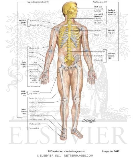
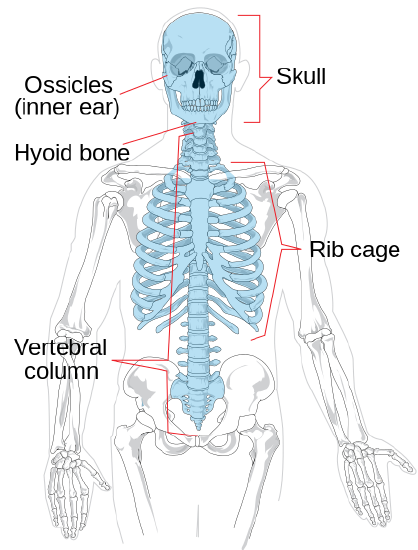
Skeletal muscles attach to and move bones by contracting and relaxing in response to voluntary messages from the nervous system. Skeletal muscle tissue is composed of long cells called muscle fibers that have a striated appearance. Muscle fibers are organized into bundles supplied by blood vessels and innervated by motor neurons. Skeletal System ANS 215 Physiology and Anatomy of Domesticated Animals I. Bones A. Cellular structures whereby the extracellular fluid environment of the cell is surrounded by a rigid, calcified frame. B. Framework of one bone, when combined with all the other bones of the body comprise the skeleton The Skeletal System • Parts of the skeletal system • Bones (skeleton) • Joints • Cartilages • Ligaments (bone to bone)(tendon=bone to muscle) • Divided into two divisions • Axial skeleton: bones of the skull, vertebral column, and rib cage • Appendicular skeleton: bones of the upper and lower limbs, shoulder and hip . Functions of Bones • Support of the body • Protection of ... Skeletal Divisions •The Axial Skeleton -Forms the longitudinal axis of the body -Has 80 bones: •The skull: -eight cranial bones -fourteen facial bones •Bones associated with the skull: -six auditory ossicles -the hyoid bone
Skeletal System Activity #4 page 7 7. Color the following areas on the diagrams of the pelvis shown below. c Pubis (C)Ileum (A) c Ischium (B) c 8. Match the bone with the correct letter from diagram at the right. _____ Cervical vertebrae _____ Coccyx ... Muscular System Tour Lab page 4 The Muscles … a front view. Deltoid. The Muscles … a back view. H. F. Use your front view and back view diagrams to label these muscles Gluteus medius Label: place the letter next to the name. Gluteus maximus . Latissimus dorsi Pectoralis major. Gastrocnemius Triceps Brachioradialis Skeletal myogenesis depends on the strict regulation of various gene subsets in order to differentiate the myogenic progenitors into myofibers. Basic helix-loop-helix (bHLH) transcription factors, MyoD, Myf5, myogenin, and MRF4 are critical to its formation. Skeletal muscles produce movement by bending the skeleton at movable joints. Muscles work in antagonistic pairs. Skeleton provides structure of body and muscles allow skeleton mobility - pull by contraction of muscle. SKELETAL SYSTEM: Functions Support & shape to body Protection of internal organs Movement in union with
(b) Explain what is meant by the expression ‘skeletal muscle pump’. Why is it important to the cardiovascular system? Question 17 What is interstitial fluid? Draw and label a diagram that shows how it is formed from plasma. Highlight your key words and write notes on your diagram to explain the process. Question 18
Have students read about the skeletal system below and take the two short answer quizzes. They can study the labeled skeleton and then try to label a whole skeleton themselves. Now they are ready to build their own labeled skeletons. 1. Print out the pdf bones of the body; hands, feet, arms, forearms legs, shins, skull, and ribcage. 2. Copy enough for each student to have a set. 3. Have them ...
finish, select different colors for each organ system and color them in. ORGAN SYSTEMS The human bodyis either studied by regions or by organs systems. This book uses the organ system approach in which individual organs (such as bones) are grouped into the larger organ system (for example, the skeletal system). Typically eleven organsystems are ...
Parts of the skeletal system include: Bones (skeleton). Joints. Cartilages. Ligaments. Divided into two divisions: 1. Axial skeleton (skull ...75 pages
System Function Diagram Major Organs Interactions- Working with Other Systems Muscular Allows for movement by contracting Cardiac muscle Smooth muscle Skeletal muscle tendons 1. w/skeletal – allow movement 2. w/digestive – allow organs to contract to push food through 3. w/respiratory – diaphragm controls breathing
SKELETAL SYSTEM CHALLENGE Label the diagram with the appropriate bone name. Created Date: 20180427155036Z ...
This textbook has been created with several goals in mind: accessibility, customization, and student engagement—all while encouraging students toward high levels of academic scholarship. Students will find that this textbook offers a strong introduction to human biology in an accessible format.
Skeleton patterns contained in this unit are just the right size for most kindergartners. Enlarge the patterns for older students.) The skeletons can be displayed on the class board as an informative but fun way to learn the bones of the body. FLASHLIGHT X-RAY After a discussion about the human skeleton, have your stu-dents experience viewing their
A diagram is a symbolic representation of information using visualization techniques. Diagrams have been used since prehistoric times on walls of caves, but became more prevalent during the Enlightenment. Sometimes, the technique uses a three-dimensional visualization which is then projected onto a two-dimensional surface. The word graph is sometimes used as a synonym for diagram.
Name:_____ Skeletal System Notes Skeletal System What are the main functions of the skeletal system? The two main functions are: • • • *There are _____ bones in the body • *Blood cells are produced in the _____ Vertebrae
Worksheet - colour in diagram of vertebrae You tube: "Cervical spine anatomy" 3:10sec. You tube: "Cervical spine" 1:43sec Spinal Discs (Intervertebral discs) The intervertebral disc in the spine is an interesting and unique structure. Its primary purpose is to act as a shock absorber between adjacent vertebrae. Spinal discs also act as ligaments that hold the vertebrae of the spine together ...
Turning to the postcranial skeleton, note which bones are part of the pectoral (shoulder) girdle and the pelvic girdle. Use the articulated skeleton in the laboratory as well as the diagrams provided to distinguish the bones of the hands (carpals, metacarpals, and phalanges) and feet (tarsals, metatarsals, and phalanges). Planes of Orientation
of the reproductive system. You can manipulate the images to see different views and isolate each ... all bold terms and diagram labels. 3 1. Scroll down to 13. ... Deselect the skeletal system by choosing. the skull icon in the list on the left.
o The SKELETAL SYSTEM Power Point slides o The PDF of the SKELETON DIAGRAM BONE IDENTIFICATION • You will need to be able to identify the following bones on a diagram, by finding the correct bone name from the word bank • All of these bones are the same ones you identified when you earned your lab stickers, so there are no surprises! SKULL: Frontal bone Parietal bone Temporal bone ...
•Parts of the skeletal system •Bones (skeleton) •Joints •Cartilages •Ligaments (bone to bone)(tendon=bone to muscle) •Divided into two divisions •Axial skeleton •Appendicular skeleton - limbs and girdle . Functions of Bones .
Unit Four - Skeletal System Page 3 Draft Copy WORKSHEET - Skeletal Tissue -KEY 1. The framework of bones and cartilage that protects our organs and allows us to move is called the skeletal system. It's functions are support, protection, movement facilitation, mineral storage, storage of blood cell - producing cells, and storage of energy. 2.
Skeletal muscles have muscle tone (remain partly contracted), which helps maintain body posture. Ongoing signals from the nervous system to the muscle cells help maintain tone and readiness for physical activity. Skeletal muscle aids heat generation. During muscle contractions, muscle cells expend much energy, most of which is converted to heat. To prevent overheating, glands in the skin ...
The skeletal system includes all of the bones, ... (PDF file). Rheumatologist. Rheumatologists are medical doctors who have additional training as internists with a sub-specialty in rheumatology. ... This diagram shows the human skeleton and identifies the major bones.
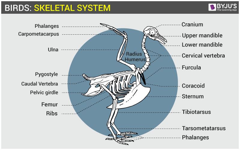
The skeletal system • The appendicular skeletal system (figure 1) consists of the shoulder girdle, skull, hip girdle, leg and arm bones. • The axial skeleton consists of the skull, vertebral column, ribs and sternum. The functions of the skeletal system are to act as a lever system, as surface area for attachment of
Parts of the Skull. The skull consists of the rounded brain case that houses the brain and the facial bones that form the upper and lower jaws, nose, ...166 pages
The Axial Skeleton. • Bones of the skull. • parts of the skull. • sinuses. • development of skull. • Vertebral column. • curves. • vertebrae & discs.38 pages
Human Anatomy & Physiology: Skeletal System; Ziser, Lecture Notes, 2010.4 2 Skeletal Anatom y each individual bone is a separate organ of the skeletal system ~ 270 bones (organs) of the Skeletal System w ith age the num ber decreases as bones fuse by adulthood the num ber is 206 (typical) even this num ber varies due to varying num bers of m ...
This tutorial will introduce you to the skeletal system. It provides information about the functions of the skeletal system, the shapes of bones, and introduces the major bones of the skeleton. The goal is to provide a basic foundation you can build upon as you learn and become more confident with Anatomy.
the bones of the human skeleton. I have concentrated on illustrations, brief descriptions and references to major sources of forensic literature. Where data is available there are summary statistics, means and standard deviations, for the dimensions of the relevant skeletal element in male and female prehistoric Aborigines.
Abstract The skeletal system is formed of bones and cartilage, which are connected by ligaments to form a framework for the remainder of the body tissues. This article, the first in a two-part series on the structure and function of the skeletal system, reviews the anatomy and physiology of bone. Understanding the structure and
Introduction to BPMN 4 Figure 2: A Segment of a Process with more Details Swimlanes Many process modeling methodologies utilizes the concept of swimlanes as a mechanism to organize activities into separate visual categories in order to illustrate different functional capabilities or
Workbook 8 | Skeletal anatomy Label the diagram with the names of the major bones M M S H R U M F F T M S C R V P C P P P T. Workbook 8 Page 9 NHS Training for AHP Support Workers Workbook 8 | Skeletal anatomy 8.6 Practical anatomy session In this session you will learn: Where some of the important bony landmarks are on the body and understand why you need to know them. Be able to identify ...
The skeleton makes up almost one-fifth of a healthy body's weight. ❖ This flexible inner framework supports all other parts and tissues, ...54 pages
1 SKELETAL SYSTEM WORKSHEET HUMAN SKELETAL SYSTEM Color the diagram below using the table as a key. Number Color Name of Bone 1. 2. 3. 4. 5. 6. 7. 8.
The skeletal system consists of 206 bones that make up the internal framework of the body, called the skeleton. The skeleton supports the body, protects internal organs, serves as a point of attachment for skeletal muscles for body movement, produces blood cells, and stores minerals. Organs Here are the primary structures that comprise the skeletal system: bones joints Word Parts Here are the ...
To be able to grow, strong bones needs lots of calcium and weight-bearing physical activity. Side of skull. (Parietal bone)2 pages









0 Response to "37 skeletal system diagram pdf"
Post a Comment